The Chin


Chapter 20
The Chin


Introduction


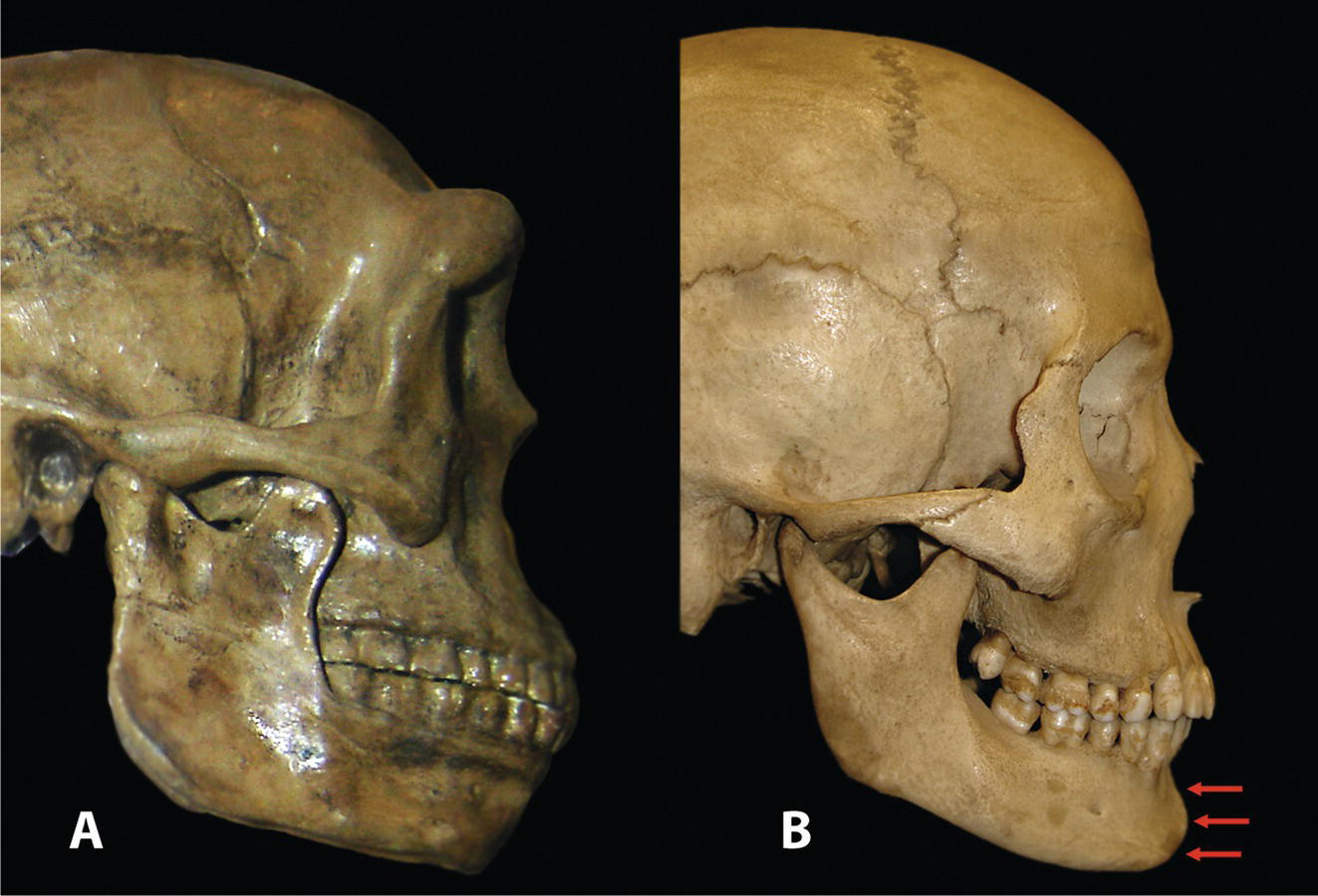
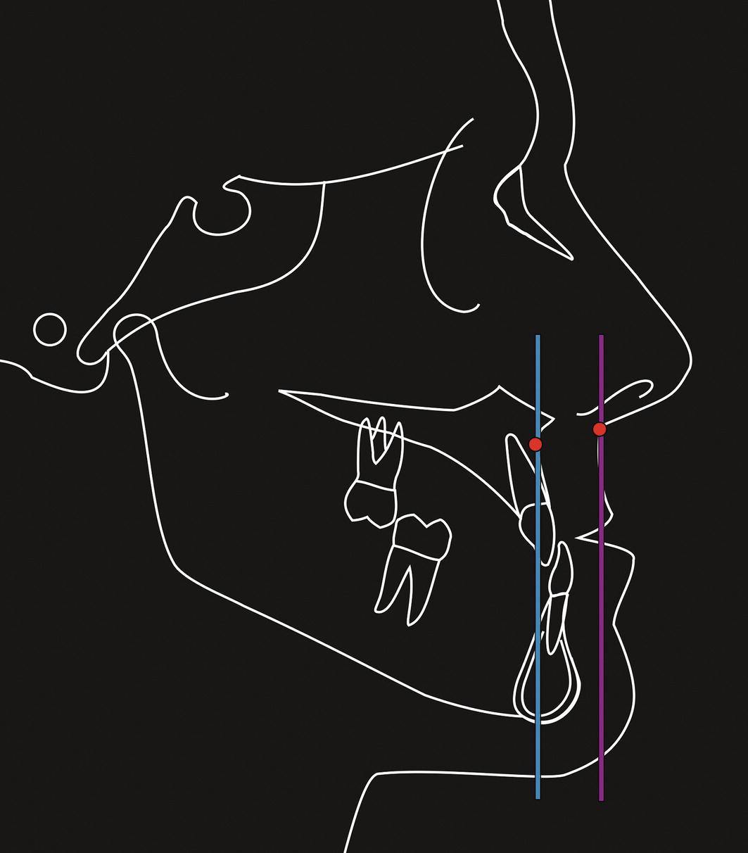
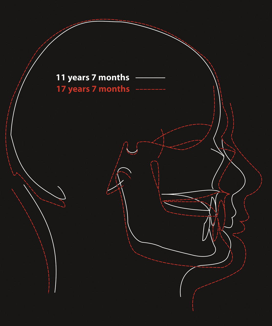
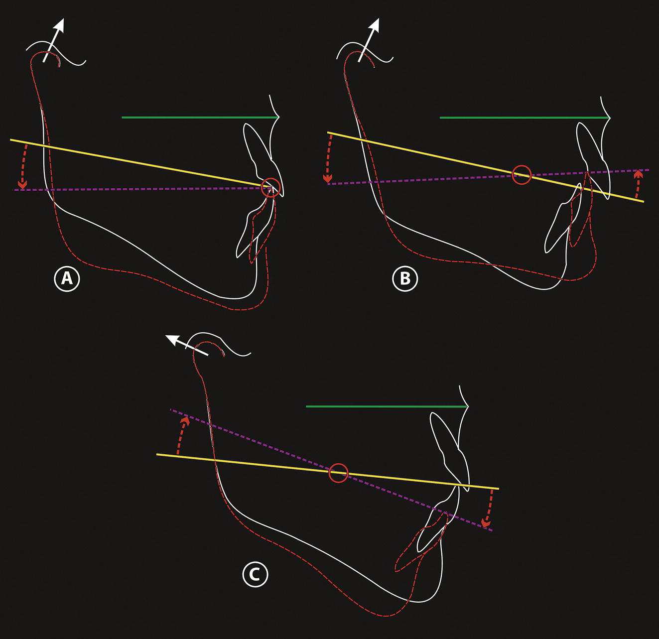
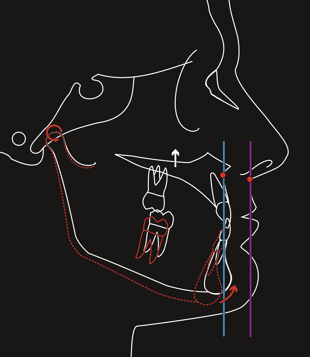
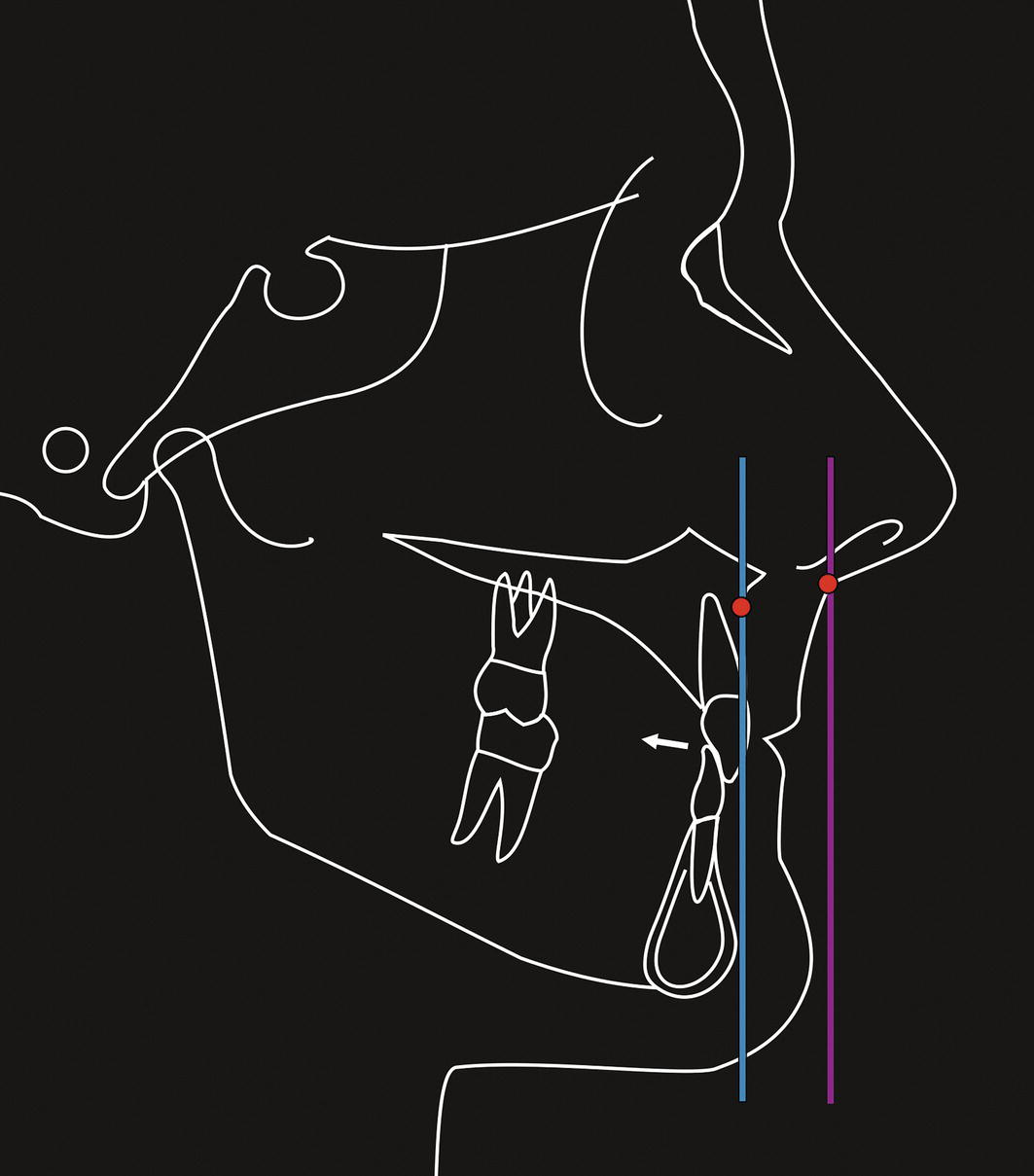
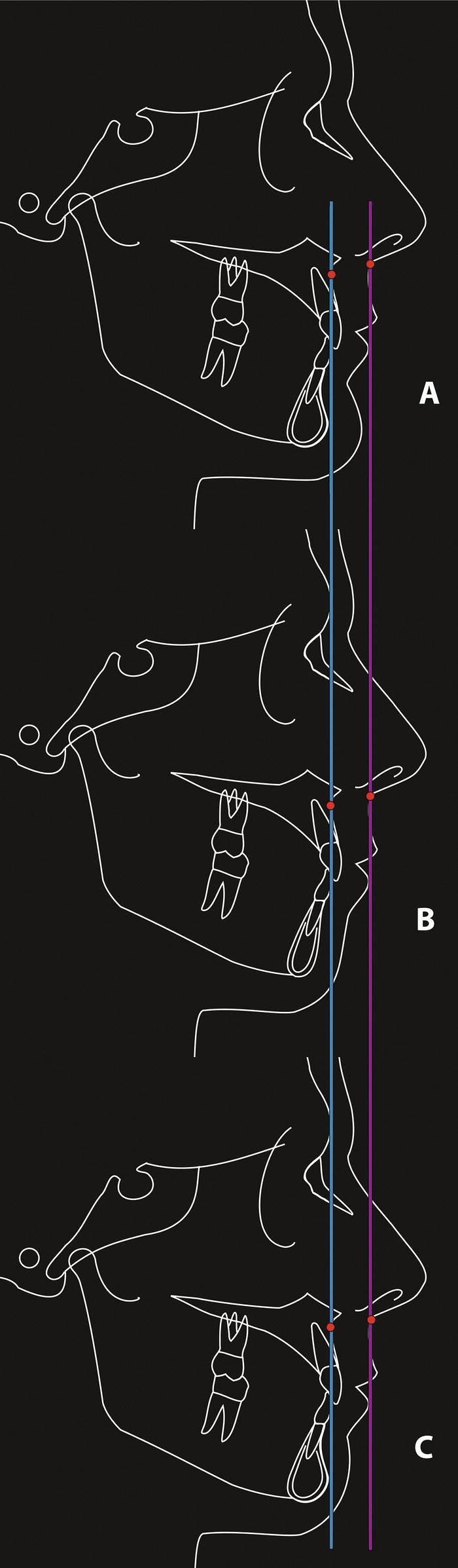
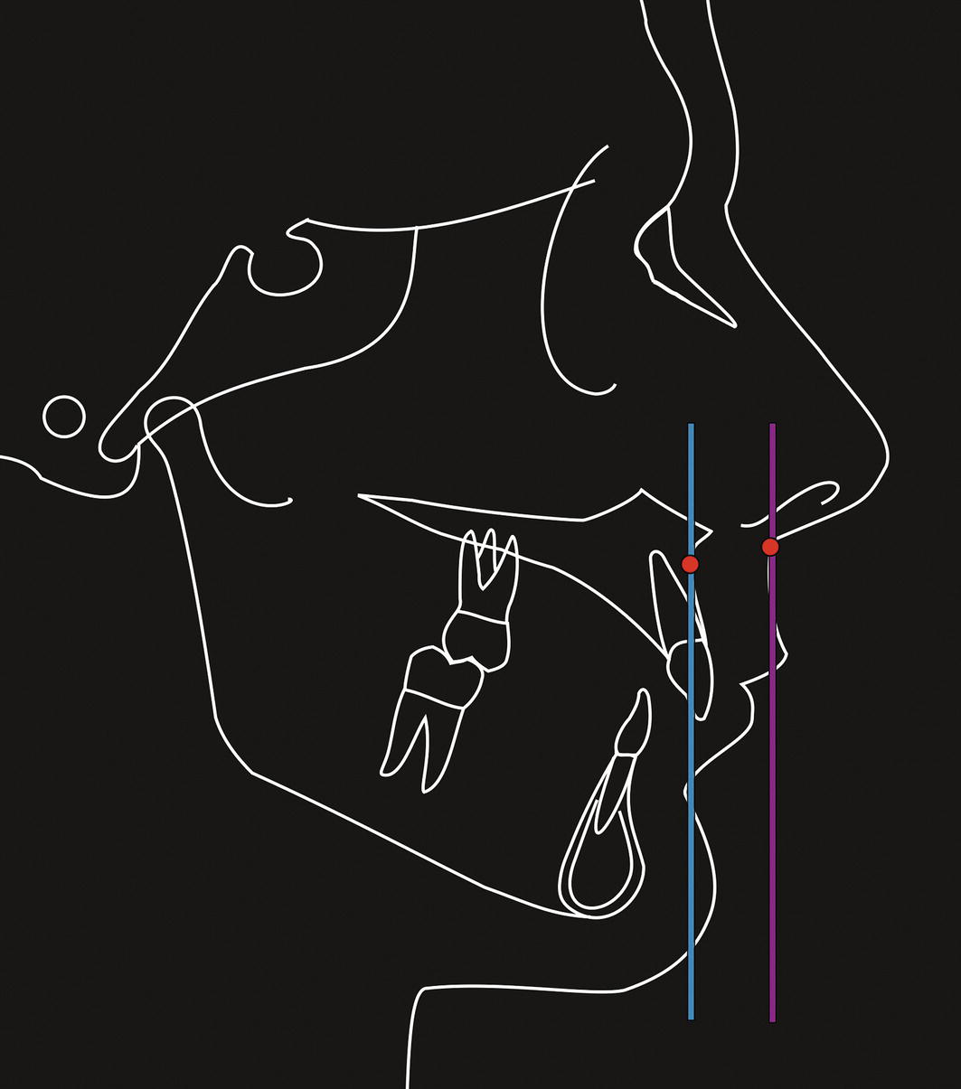
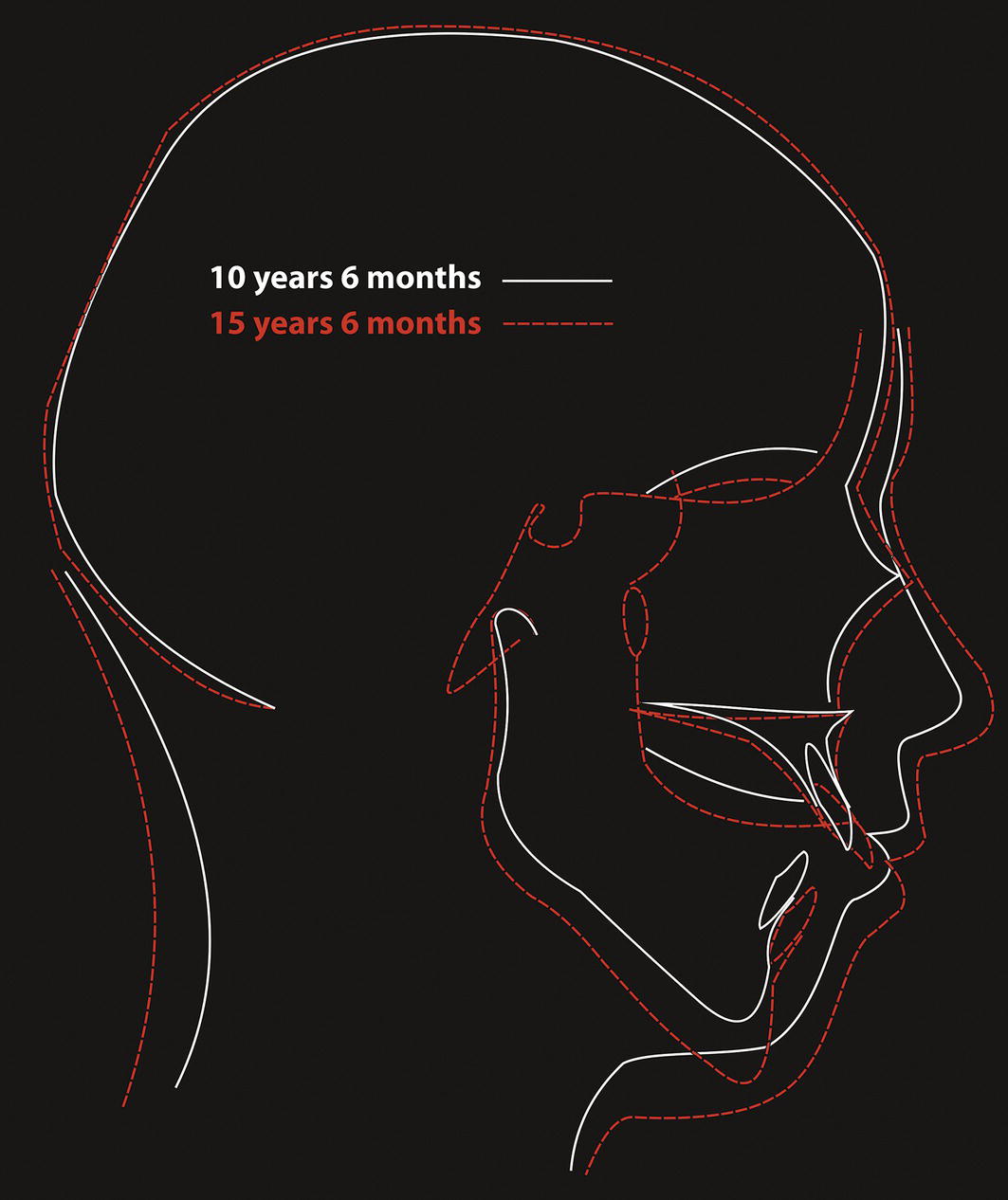
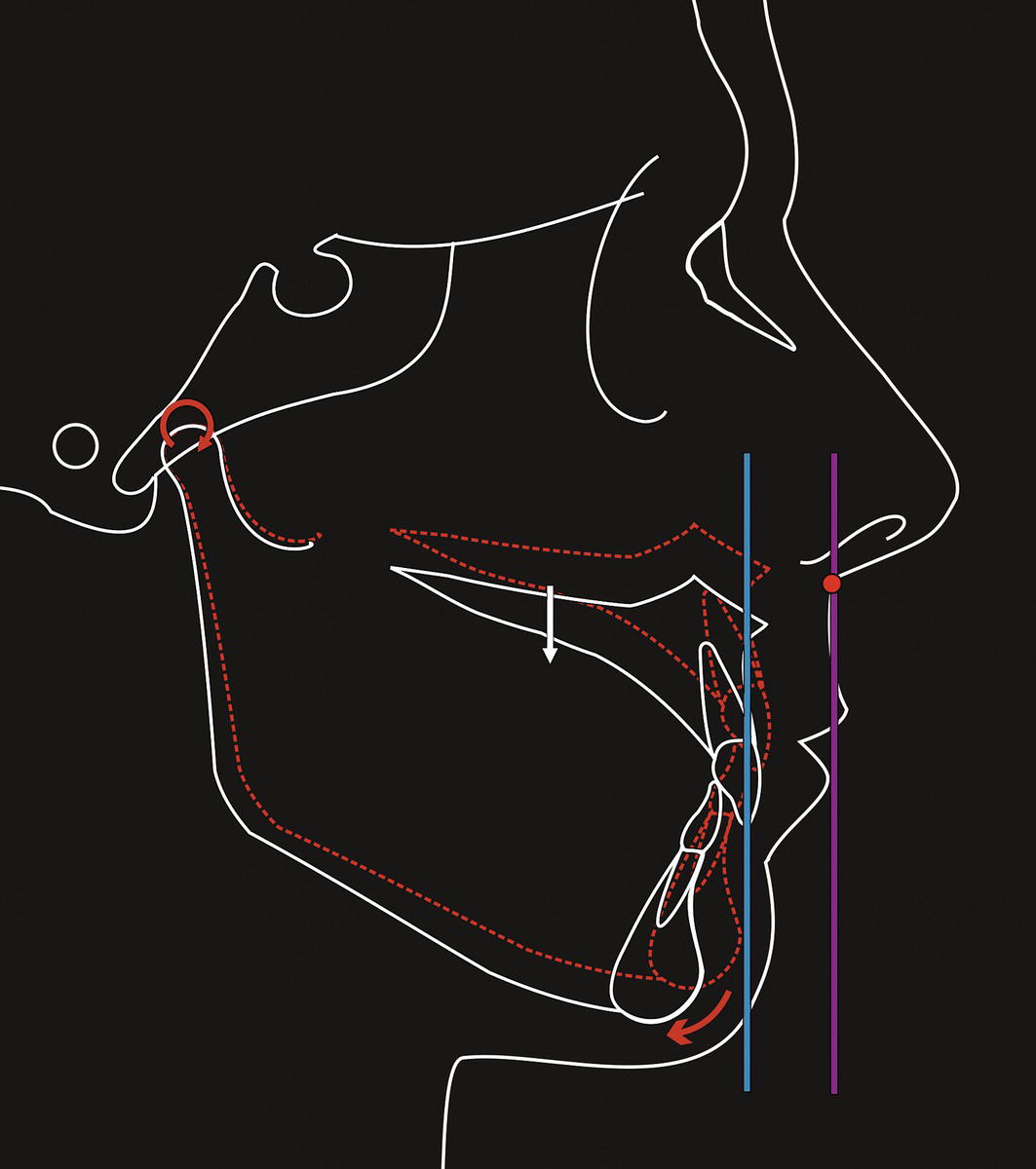
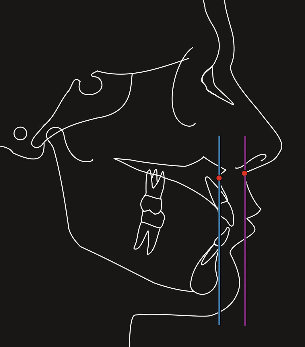
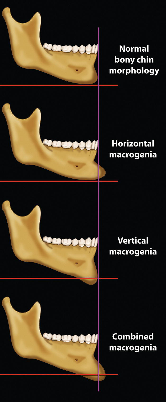
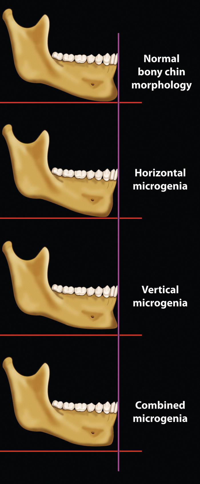
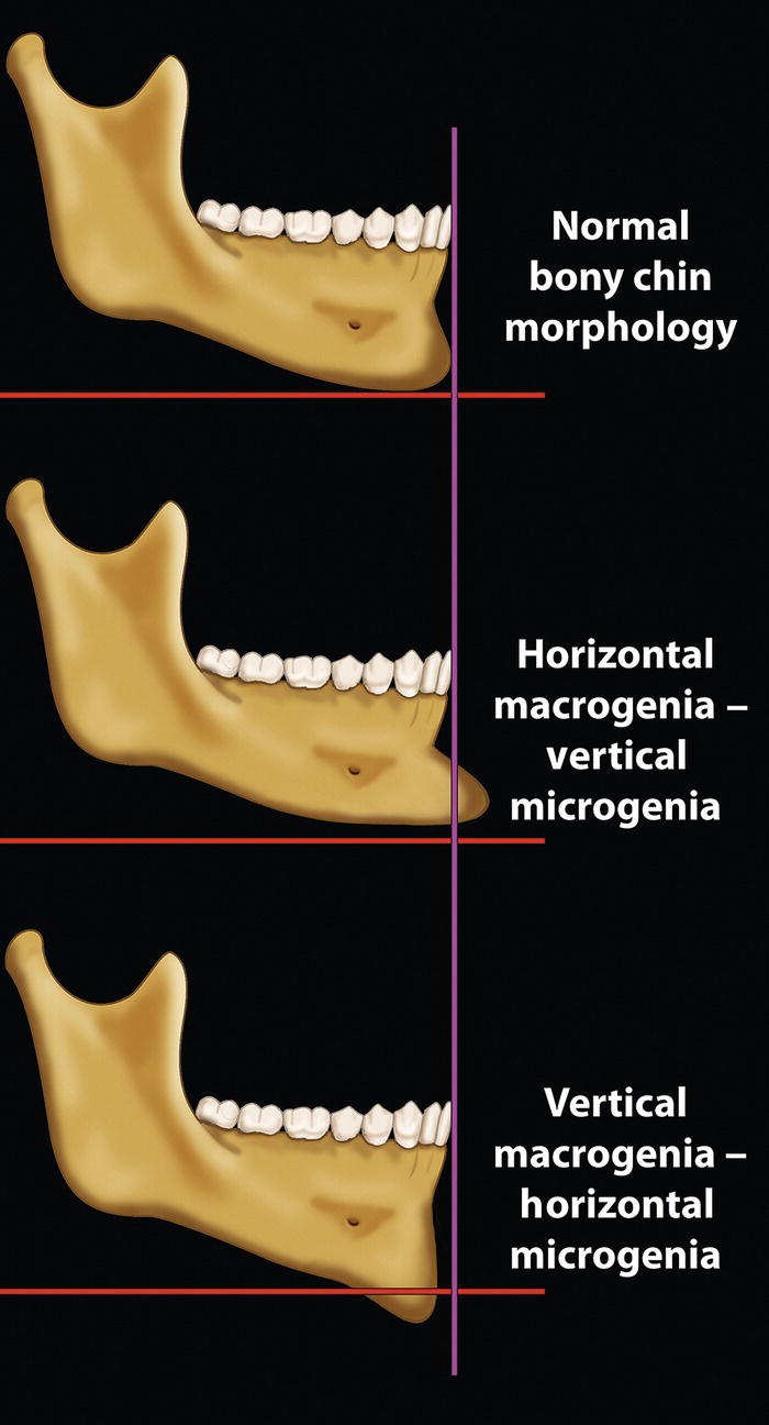
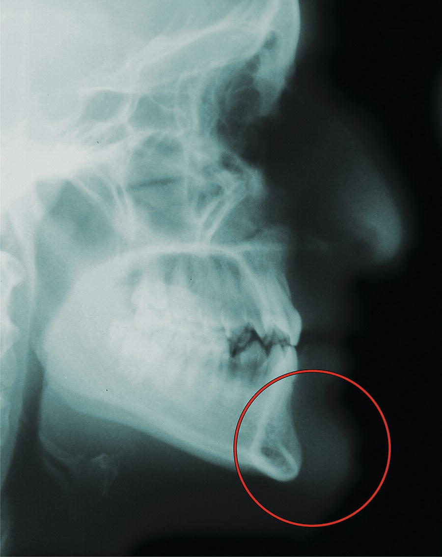
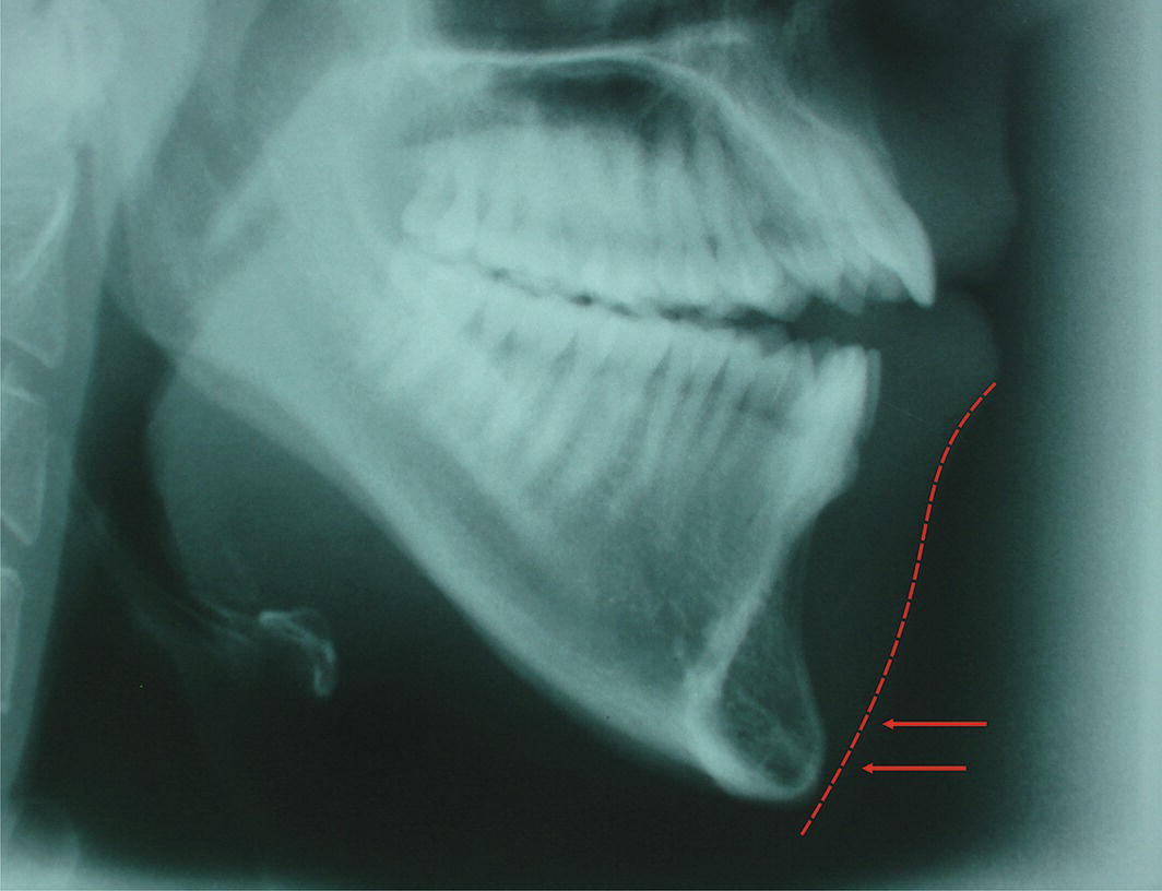
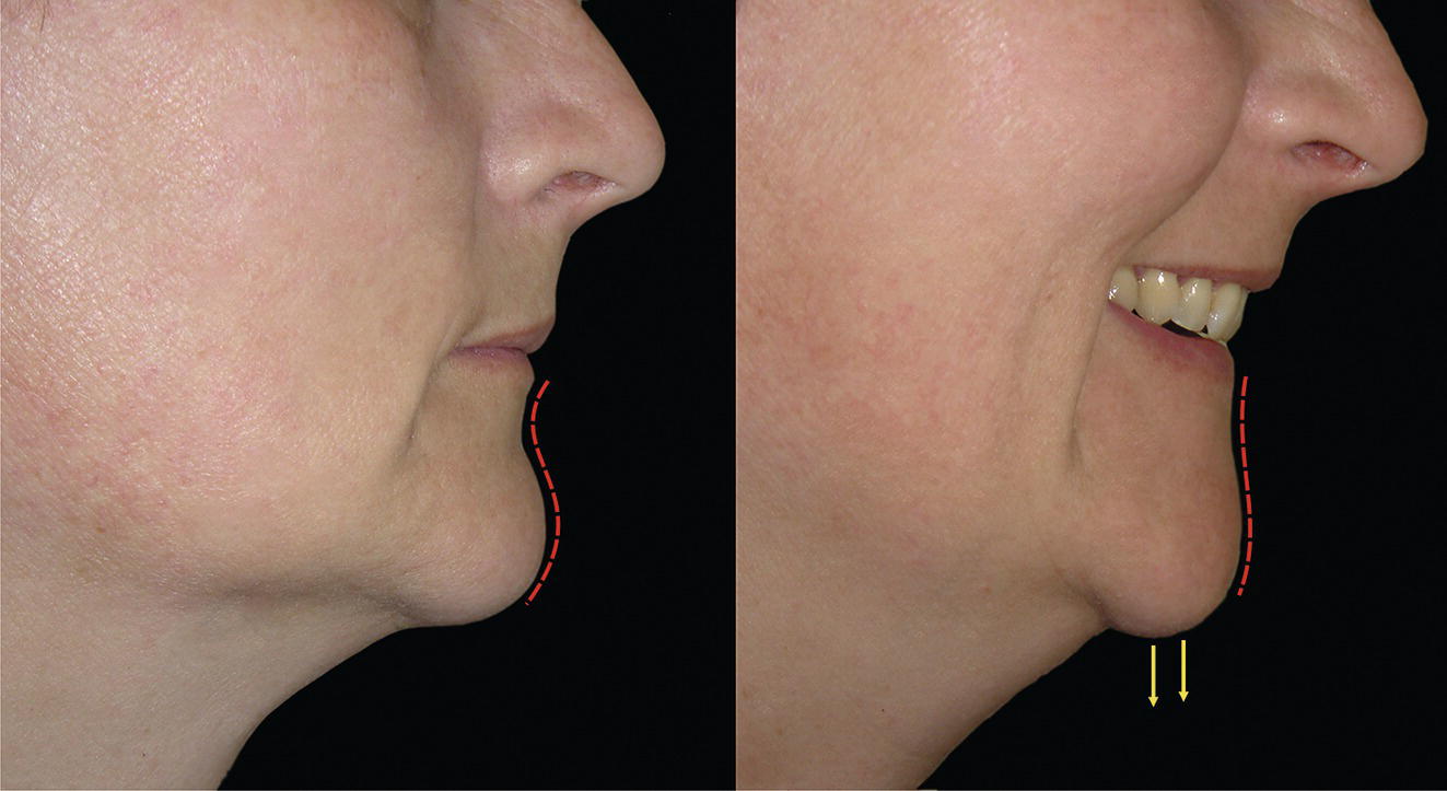
‘Thou whose features clearly beaming make the moon


of Beauty bright,


Thou whose chin contains a well‐pit which to


Loveliness gives light’.


Hafez (1325–90)


Persian poet‐philosopher1


The chin is unique to humans. The human face as we know it originated with the Homo sapiens in Africa, approximately 130 000 years ago. Even Neanderthals retained a modest muzzle, whereas the need for a projecting mouth all but disappeared in modern humans. This flattening of the face and retrusion of the orodental complex coincided with the appearance and projection of the bony chin, perhaps a remnant of the now obsolete muzzle (Figure 20.1).


The morphology of the chin has a substantial influence on the perceived attractiveness of the face. In profile view, in particular, the chin establishes much of the character of the lower face (Figure 20.2). In fact, the prominence of the chin is one of the facial characteristics that society tends to associate with an individual’s personality. Individuals, particularly men, with a deficient chin may be viewed as ‘weak’, whereas a prominent chin is often described as a ‘strong’ chin, implying strength of personality.


The history of aesthetic chin surgery has been described comprehensively elsewhere.2Otto Hofer (1942) is likely to have been the first surgeon to propose a horizontal osteotomy of the mandibular symphysis and sliding advancement of the inferior fragment.3 The procedure was performed from an external approach and the ‘patient’ seems to have been a cadaver. The first report of aesthetic chin surgery in a living patient seems to be that of Sir Harold Gillies and Ralph Millard Jr (1957).4 The procedure was performed through an external approach, with the inferior fragment being advanced and positioned superiorly, anterior to the upper mandibular fragment; this procedure is now termed a ‘jumping genioplasty’. Richard Trauner and Hugo Obwegeser (1957) later reported aesthetic chin surgery through an intraoral approach, and coined the term ‘genioplasty’.5John Converse and Donald Wood‐Smith (1964) reported their results through an intraoral approach, referring to the procedure as a ‘horizontal osteotomy of the mandible’.6Thomas Hohl and Bruce Epker (1976) discussed vertical reduction of chin height by wedge ostectomy.7Kevin McBride and William Bell (1980) discussed increasing the height of the chin with autogenous interpositional bone graft.8


Anatomy


Anatomically the chin is the area below the mentolabial fold, although separating the chin from the lower lip in patients with a poorly defined mentolabial fold can be difficult, particularly in frontal view. The anatomy, morphology and aesthetics of the lower lip, mentolabial fold and chin are intimately related. The chin consists of the bony anterior projection of the mandible, called the mandibular or mental symphysis, and its overlying soft tissue chin pad.

An image features a pair of human skulls representing different species. The first skull belongs to Homo erectus, an ancient hominid that lived over a million years ago, exhibiting distinct cranioskeletal morphology. The second skull is from modern Homo sapiens, showcasing key differences such as a larger cranium, taller forehead, and smaller teeth. These variations highlight the evolutionary changes between the two species.

Figure 20.1 (A) Cranioskeletal morphology of Homo erectus, a species of hominid (of the human family) that walked upright and lived more than one million years ago.


(With kind permission, © Natural History Museum, London.)


(B) Cranioskeletal morphology of modern human (modern Homo sapiens) demonstrates the differences between the two species: Homo sapiens has a larger cranium, a taller forehead, reduced prominence of the brow ridge, reduced mandibular length, reduced bimaxillary protrusion, smaller teeth and a pronounced bony chin.

A detailed study focusing on the chin and its significance in defining the character of the lower face, especially from a profile perspective. It includes elements of a human face, highlighting attributes such as the forehead and cheek. The context is tied to an artistic study of the depiction of Leda.

Figure 20.2 The chin establishes much of the character of the lower face, particularly in profile view.


(Detail, Study for the Face of Leda, Michelangelo, c. 1530, Casa Buonarroti, Florence.)


Terminology


The ability to accurately describe and thereby classify facial deformities is essential, and a prerequisite to correct diagnosis, leading to correct treatment planning. An important step towards classification is therefore to define the clinical terminology based on etymology. The terminology used should reflect the underlying aetiology.


  • Genio‐ (Greek geneoin: chin).
  • Mento‐ (Latin mentum: chin).
  • Pogo‐, pogono‐ (Greek: beard).
  • Macro‐ (Greek macros: large or abnormally large).
  • Micro‐ (Greek micros: small or abnormally small).
  • Pro‐ (prefix Greek, Latin: forward).
  • Retro‐ (prefix Latin: backward or behind in position).

Chin excess and chin deficiency


The terms ‘chin excess’ and ‘chin deficiency’ may be used to describe chin deformities in the sagittal or vertical plane.


Progenia (sagittal chin excess)


Progenia is a term used to indicate that the sagittal position of soft tissue pogonion (Pog′, the most prominent point on the soft tissue contour of the chin, in the midsagittal plane) is protrusive (too far forward) in relation to the rest of the craniofacial complex. This may be due to a relatively larger size (macrogenia) and/or a more anterior position of the chin.


Progenia is a non‐specific term describing the sagittal position of the chin, not the underlying aetiology. Progenia may be classified as primary, secondary or ‘relative’.


Primary progenia (primary sagittal chin excess): This is a structural problem, describing a morphological deformity of the chin area. An alternative and preferred term is ‘sagittal chin excess’. The aetiology of sagittal chin excess may be due to one or a combination of the following:

A diagram focusing on different types of chin prominence. It includes references to normal chin prominence and variations such as horizontal often microglia and thickening of the soft tissue chin pad. The overall presentation suggests a technical or medical illustration, possibly used for educational purposes related to facial anatomy.

Figure 20.3 (A) Normal chin prominence. (B) Horizontal osseous microgenia. (C) Increased thickness of soft tissue chin pad.

A diagram that illustrates a sagittal chin excess resulting from mandibular excess. It highlights the normal morphology of the chin in relation to a prominent mandible. The content suggests a focus on anatomical relationships and variations.

Figure 20.4 Sagittal chin excess secondary to mandibular excess (i.e. normal chin morphology on a prominent mandible).


  • Horizontal osseous macrogenia (Figure 20.3B)
  • Increased thickness of soft tissue chin pad (Figure 20.3C)
  • Any combination of the above.

Secondary progenia (secondary sagittal chin excess): This is a positional problem of the mandible, describing the forward position of the chin as a result of an excessively forward position of the mandible, i.e. the morphology of the chin is normal; the abnormal position of the chin is secondary to the abnormal position of the mandible, with the normal chin being carried forward on the mandible. The aetiology of secondary progenia may be due to one or a combination of the following:


  • Mandibular excess (prognathism/macrognathism) (Figure 20.4).
  • Hypodivergent facial growth pattern (Figure 20.5): Alternative terms are ‘horizontal facial growth pattern’, ‘low angle’ patient (referring to the reduced mandibular plane angle), ‘short face deformity’ or ‘short face syndrome’. If the hypodivergent growth is the primary aetiology to a deep incisor overbite, the term ‘skeletal deep bite’ may be used. An element of vertical maxillary deficiency (VMD) often coincides with mild mandibular retrognathia, leading to a reduced lower anterior face height (LAFH), a reduced mandibular plane angle and mandibular overclosure. This leads to an increased sagittal projection of the chin.9 Such hypodivergent facial growth patterns will tend to be associated with an anterior pattern of mandibular growth rotation.
    A diagram that illustrates the facial growth patterns associated with a short face type. It specifically highlights horizontal and hypo divergent growth leading to an increased sagittal projection of the chin over two age milestones: 11 years 7 months and 17 years 7 months.

    Figure 20.5 Horizontal, hypodivergent (short face type) facial growth pattern, leading to an increased sagittal projection of the chin.


    (Modified from Björk9 with permission from Informa Healthcare, Taylor and Francis Group.)


  • Anterior (forward) mandibular growth rotation (Figure 20.6A,B): Growth rotations of the mandible occur when there is a discrepancy in the amounts of growth in anterior and posterior facial heights. Anterior rotational growth of the mandible affects sagittal chin position by moving the chin upward and forward, increasing the sagittal projection of the chin. Implant studies by Björk placed the centre of rotation at either the incisal edges of the mandibular incisors or at the premolars.10
  • Anterior mandibular autorotation around the condylar hinge axis (Figure 20.7). This may be due to:

    • Loss of the dentition and associated alveolar processes leads to mandibular overclosure, with the chin rotating closer towards the nose. This has a tendency to occur with ageing.
    • Maxillary impaction surgery: The mandible will thereby autorotate forward and upward around the condylar hinge axis.

  • Anterior mandibular displacement: In the presence of a mild Class III incisor relationship, patients with an uncomfortable edge‐to‐edge incisor occlusion may displace and posture the mandible anteriorly in order to achieve maximum intercuspation of the posterior teeth (see Figure 19.16).
  • Any combination of the above.

‘Relative’ progenia (relative sagittal chin excess): The chin appears protrusive (too far forward) due to lower labial or bilabial retrusion, i.e. the lower lip or both lips being positioned too far back in relation to the craniofacial complex (Figure 20.8).


Retrogenia (sagittal chin deficiency)


Retrogenia is a term used to indicate that the sagittal position of soft tissue pogonion (Pog’) is retrusive (too far back) in relation to the rest of the craniofacial complex. This may be due to a relatively smaller size (microgenia) and/or a more posterior position of the chin.


Retrogenia is a non‐specific term describing the position of the chin, not the underlying aetiology. Retrogenia may be classified as primary, secondary or ‘relative’.


Primary retrogenia (primary sagittal chin deficiency): This is a structural problem, describing a morphological deformity of the chin area. An alternative and preferred term is ‘sagittal chin deficiency’. The aetiology of sagittal chin deficiency may be due to one or a combination of the following:


  • Horizontal osseous microgenia (Figure 20.9B)
  • Reduced thickness of soft tissue chin pad (Figure 20.9C)
  • Any combination of the above.

Secondary retrogenia (secondary sagittal chin deficiency): This is a positional problem of the mandible, describing the retrusive position of the chin as a result of an excessively retropositioned mandible, i.e. the morphology of the chin is normal; the abnormal position of the chin is secondary to the abnormal position of the mandible. The aetiology of secondary retrogenia may be due to one or a combination of the following:


  • Mandibular deficiency (retrognathia/micrognathia) (Figure 20.10).
  • Hyperdivergent facial growth pattern (Figure 20.11): Alternative terms are ‘vertical facial growth pattern’, ‘high angle’ patient (referring to the increased mandibular plane angle), ‘long/tall face deformity’ or ‘long face syndrome’. If the hyperdivergent growth is the primary aetiology to an anterior open bite, the terms ‘skeletal open bite’ or ‘apertognathia’ may be used. The maxilla rotates downward and backward (posterior VME), as does the mandible during growth. This leads to reduced sagittal projection of the chin.9 Such a vertical facial growth pattern will tend to be associated with a posterior pattern of mandibular growth rotation.
  • Posterior (backward) mandibular growth rotation (Figure 20.6C): Posterior rotational growth of the mandible affects sagittal chin position by moving the chin downward and backward, effectively reducing the sagittal projection. Implant studies by Björk place the centre of rotation at the occluding molars.10
  • Posterior mandibular autorotation around the condylar hinge axis (Figure 20.12). This may be due to maxillary inferior repositioning – the mandible autorotates downward and backward.
  • Any combination of the above.
A diagram depicting types of mandibular growth rotation. It includes three variations: (A) anterior growth with the centre at the incisal edges, resulting in upward and forward condylar growth; (B) anterior growth with the centre at the premolars, also moving the chin upward and forward; and (C) posterior growth that moves the chin downward and backwards. The diagram highlights how these rotations affect chin projection.

Figure 20.6 Types of mandibular growth rotation. (A) Anterior (forward) mandibular growth rotation with the centre of rotation at the incisal edges of mandibular incisors; the direction of condylar growth is upward and forward. (B) Anterior mandibular growth rotation with the centre of rotation at the premolars. In both cases, the chin moves upward and forward, increasing chin projection. (C) Posterior (backward) mandibular growth rotation moving the chin downward and backward, effectively reducing the sagittal projection.


(Modified from Björk and Skieller10/with permission of Elsevier.)

A diagrammatic representation of the concept of anterior mandibular autorotation around the condylar hinge axis. This process is shown to increase sagittal chin projection. The diagram likely includes various sketches and graphics to explain this anatomical movement clearly.

Figure 20.7 Anterior mandibular autorotation around the condylar hinge axis will increase sagittal chin projection.

A diagrammatic representation of relative sagittal chin excess, highlighting how the chin can appear protrusive due to lower labial or bilabial retrusion. It includes labelled elements to convey anatomical relationships effectively. The drawing serves as an informative visual aid in understanding this specific facial structure.

Figure 20.8 Relative sagittal chin excess, with the chin appearing protrusive (too far forward) due to lower labial or bilabial retrusion.

A diagram featuring three labelled parts related to chin prominence. It includes references to normal chin prominence, a horizontal configuration, and reduced thickness of the soft tissue chin pad. The visual elements include sketches and handwritten notes that enhance the understanding of these concepts.

Figure 20.9 (A) Normal chin prominence. (B) Horizontal osseous microgenia. (C) Reduced thickness of soft tissue chin pad.


‘Relative’ retrogenia (relative sagittal chin deficiency): The chin appears retrusive (too far back) due to lower labial or bilabial protrusion, i.e. the lower lip or both lips being positioned too far forward in relation to the craniofacial complex (Figure 20.13).

A diagram illustrating sagittal chin deficiency that occurs due to mandibular deficiency. It shows a comparison of normal chin morphology associated with a small or retropositioned mandible. The visual aids are designed to clarify the anatomical aspects involved in this condition.

Figure 20.10 Sagittal chin deficiency secondary to mandibular deficiency (i.e. normal chin morphology on a small or retropositioned mandible).


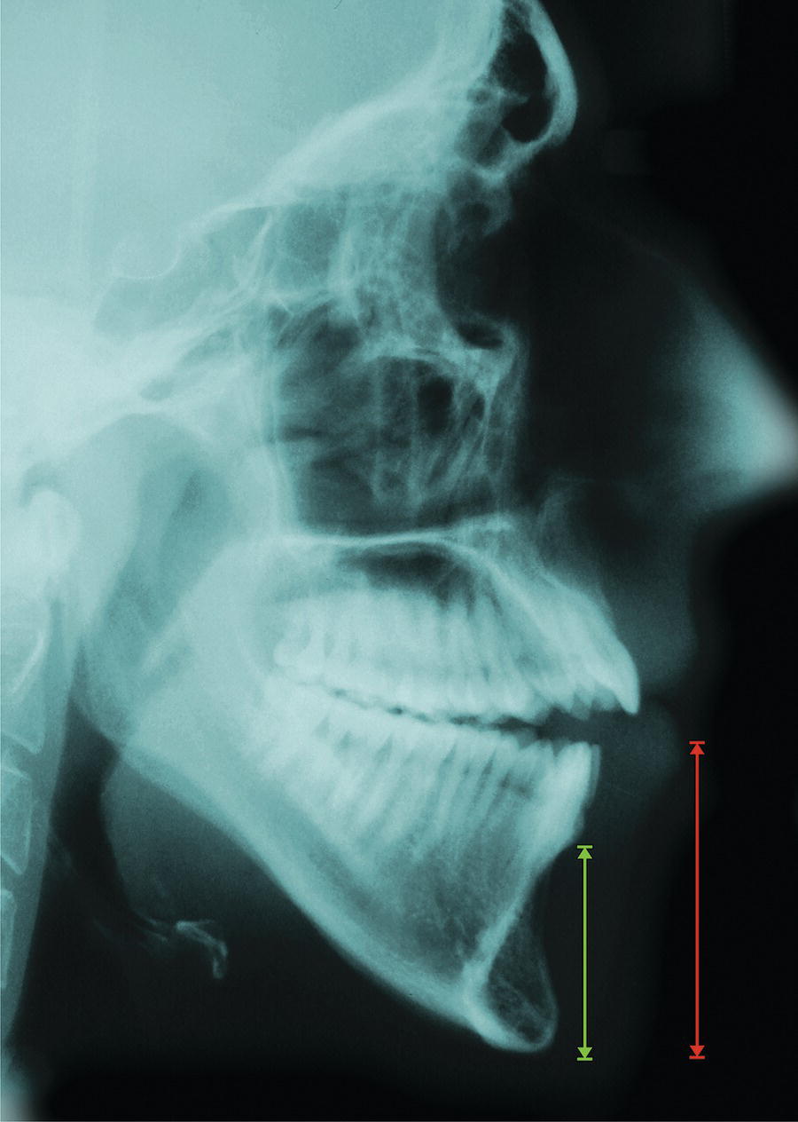
Vertical chin excess (VCE)


This term describes an increase in vertical skeletal chin height (Figure 20.15).

A diagram illustrates the vertical, hyper divergent facial growth pattern, specifically categorized as the 'tall face' type. It highlights how this growth pattern can lead to a reduced sagittal projection of the chin over a period spanning from 10 years and 6 months to 15 years and 6 months.

Figure 20.11 Vertical, hyperdivergent (‘tall face’ type) facial growth pattern, leading to a reduced sagittal projection of the chin.


(Modified from Björk9 with permission from Informa Healthcare, Taylor and Francis Group.)

A diagram illustrates the concept of posterior mandibular autorotation around the condylar hinge axis. It highlights how this movement reduces sagittal chin projection.

Figure 20.12 Posterior mandibular autorotation around the condylar hinge axis reduces sagittal chin projection.


Vertical chin deficiency (VCD)


This term describes a reduction in vertical skeletal chin height.


Classification of chin deformities
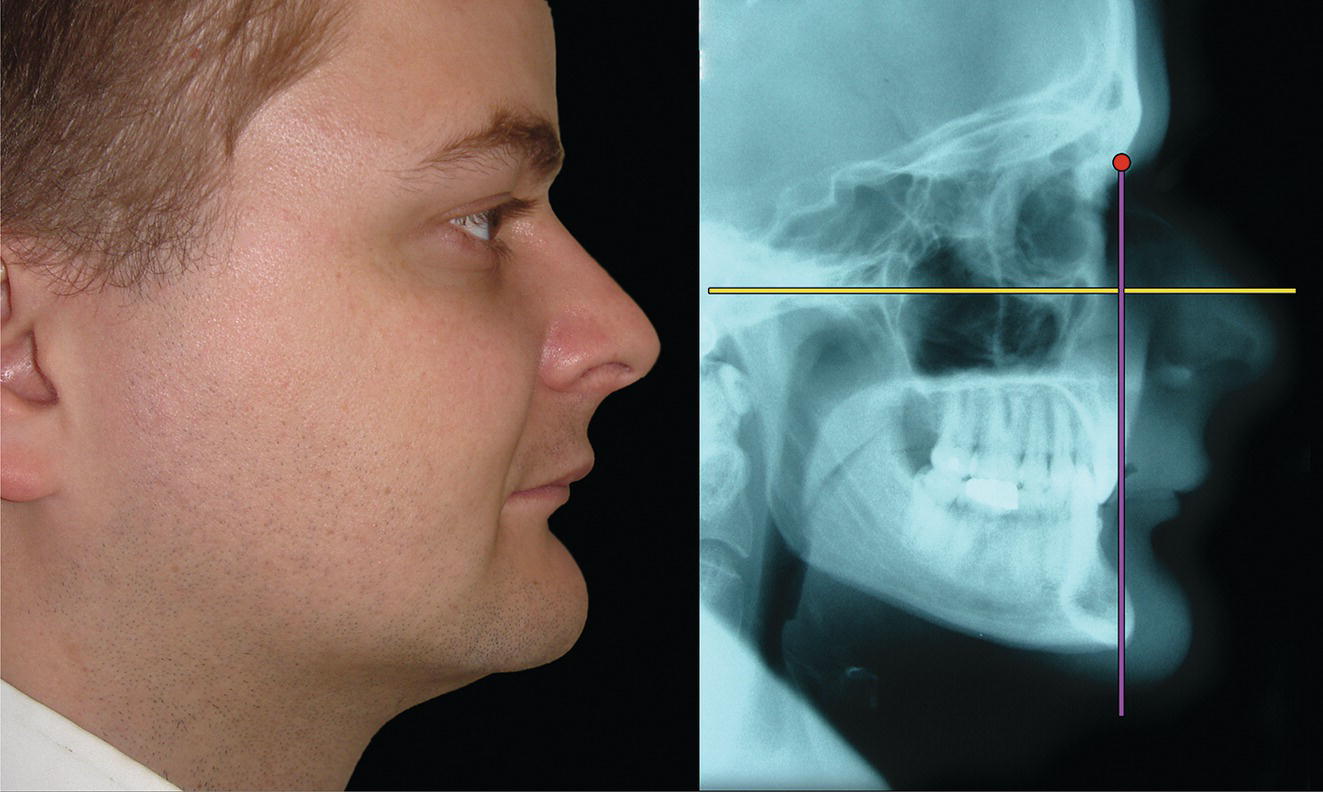

Guyuron et al. (1995)11 proposed a classification of chin deformities to be used as a guide to surgical treatment planning. They identified seven categories of chin deformities from 684 patients with normal dental occlusions. A modification of this classification is presented below:


Skeletal chin (mandibular symphysis):


  • Macrogenia (Figure 20.16):

    • Horizontal
    • Vertical
    • Combined horizontal and vertical.

  • Microgenia (Figure 20.17):

    • Horizontal
    • Vertical
    • Combined horizontal and vertical.

  • Combined (Figure 20.18):
    A diagram of a sagittal chin deficiency. It includes relevant information about relative sagittal chin deficiency, illustrating how the chin appears retrusive due to lower labial or bilabial protrusion.

    Figure 20.13 Relative sagittal chin deficiency, with the chin appearing retrusive (too far back) due to lower labial or bilabial protrusion.

    An image depicts a sagittal mandibular deficiency along with horizontal macrogenia, commonly referred to as sagittal chin excess. This is likely part of a medical imaging study related to radiology. The content also involves terms associated with medical radiography and x-ray films, indicating its clinical relevance.

    Figure 20.14 Sagittal mandibular deficiency combined with horizontal macrogenia (sagittal chin excess).

    A xray describes a condition involving vertical chin excess, indicating that the lower anterior dentoalveolar and chin height have increased primarily due to an increase in skeletal chin height. The content is tagged with terms relevant to medical imaging, such as x-ray film and radiology.

    Figure 20.15 Vertical chin excess – lower anterior dentoalveolar and chin height is increased primarily as a result of an increase in skeletal chin height.


    • Horizontal macrogenia with vertical microgenia
    • Horizontal microgenia with vertical macrogenia.

  • Asymmetric chin
A schematic diagram depicts different types of skeletal chin excess, specifically focusing on macrogenia. It details three variations: horizontal macrogenia, vertical macrogenia, and combined macrogenia. Additionally, it includes information about the normal bony chin morphology and a specific reference to "16 Macrogenla."

Figure 20.16 Macrogenia – types of skeletal chin excess.

A diagram presents information about microgenia, a condition characterized by various types of skeletal chin deficiency. It includes descriptions of normal chin morphology as well as specific types: horizontal microgenia, vertical microgenia, and combined microgenia. Each type is defined in terms of its structural characteristics.

Figure 20.17 Microgenia – types of skeletal chin deficiency.

A diagram related to chin morphology, detailing different types of chin deformities, including combined skeletal chin deformities. It describes two specific conditions: macrogenia and microgenia, both in horizontal and vertical orientations. The design contains textual information regarding these morphological characteristics.

Figure 20.18 Combined skeletal chin deformity.


Soft tissue chin pad:


  • Increased thickness of soft tissue chin pad (Figure 20.19).
  • Reduced thickness of soft tissue chin pad (Figure 20.20).
  • Ptosis of soft tissue chin pad (witch’s chin deformity) (Figure 20.21).

Mandibular rotation:


  • Chin deficiency due to posterior mandibular rotation, secondary to posterior VME in a vertical facial growth pattern and/or posterior (backward) mandibular growth rotation.
  • Chin excess due to anterior mandibular rotation, secondary to VMD in a horizontal facial growth pattern and/or anterior (forward) mandibular growth rotation.

Mandibular position and/or size:


  • Chin deficiency secondary to mandibular deficiency (retrognathia or micrognathia).
    An x-ray of a person's chin area, showing increased thickness of the soft tissue chin pad, while the bony chin appears relatively underdeveloped. It falls under medical imaging and radiology categories.

    Figure 20.19 Increased thickness of soft tissue chin pad (bony chin is relatively underdeveloped).


  • Chin excess secondary to mandibular excess (prognathia or macrognathia).
  • Chin asymmetry secondary to mandibular asymmetry.

Clinical evaluation


The morphology of the mandible tends to be relatively consistent depending on the presenting facial deformity, e.g. a Class II, Class III, tall face or short face individual tends to have a ‘typical’ mandibular shape. The chin is part of the mandible; however, the morphology of the osseous chin, or mandibular symphysis, and the overlying soft tissue chin pad is highly variable in all three planes of space, even with the same basic types of facial deformities. Therefore, the chin must be evaluated as an independent facial aesthetic subunit.


When evaluating the chin, the following patient positioning guidelines should be observed:


  • Natural head position (NHP): The patient must be examined in NHP. It is not uncommon for patients to develop a compensatory head posture in order to minimize the aesthetic impact of their facial appearance, e.g. Class II patients may tilt their heads up to increase their chin prominence, Class III patients may tilt their heads down to reduce chin prominence.
    An x-ray film illustrates a specific medical condition, showing the reduced thickness of the soft tissue chin pad with noticeable mentalis hypertrophy at a higher level to the chin. The content falls under medical imaging and radiology categories.

    Figure 20.20 Reduced thickness of soft tissue chin pad (mentalis hypertrophy is evident at a higher level to the chin).

    Two photographs presenting data related to facial anatomy. It specifically references ptosis of the soft tissue chin pad, comparing its appearance in repose and during animation.

    Figure 20.21 Ptosis of the soft tissue chin pad in repose and in animation.


  • Mandibular rest position: It is important that the mandible is not postured or overclosed:

    • Mandible postured: Patients with Class II skeletal relationships may have a tendency to posture the mandible anteriorly. Some patients become so used to these postured positions, which may be up to 10 mm, that it is possible for the clinician to miss the postured position.
    • Mandible overclosed: This is particularly common in patients with maxillary hypoplasia.

  • Soft tissues in repose: The facial soft tissues must be at rest, in particular avoiding any lip strain to achieve a lip seal.
Nov 8, 2025 | Posted by in Aesthetic plastic surgery | Comments Off on The Chin

Full access? Get Clinical Tree

Get Clinical Tree app for offline access