Dermatitis is a frequent cause for referral to the pediatric dermatologist. In this article, a brief overview is given of common childhood dermatoses as well as some rarer dermatoses that may give the clinician cause for concern. Widespread scaling and erythema, described as erythroderma, are a cause of frustration for patients, families, and their physician(s). Both unusual and common skin disorders can present in this fashion. Just as recognizing common dermatoses is important, it is also important to recognize when a dermatitis fails to fit the common pattern and may prompt further investigation.
Key points
- •
Dermatitis may be localized to the diaper area or to periorificial areas in children, giving possible clues to the cause.
- •
The more common causes of widespread dermatitis include atopic dermatitis, infantile seborrheic dermatitis, and psoriasis.
- •
Rarer causes of widespread recalcitrant dermatitis are many and include ichthyoses, immunodeficiency, metabolic, and neoplastic disorders.
- •
Recalcitrant dermatitis may be difficult to treat and may be a harbinger of an alternative and more worrisome diagnosis prompting further workup.
Dermatitis is a frequent cause for referral to the pediatric dermatologist ( Box 1 ). In this article, a brief overview is given of common childhood dermatoses, as well as some rarer dermatoses that may give the clinician cause for concern. Widespread scaling and erythema, described as erythroderma, are a cause for frustration for patients, families, and their physician(s). Both unusual and common skin disorders can present in this fashion. Just as recognizing common dermatoses is important, it is also important to recognize when a dermatitis fails to fit the common pattern and may prompt further investigation.
- •
Atopic dermatitis
- •
Seborrheic dermatitis
- •
Psoriasis
- •
Contact dermatitis
- •
Infectious diseases
- •
Drug eruptions
- •
Inherited disorders
Localized dermatitis
Dermatitis may be localized to the diaper area or to periorificial areas in children, giving possible clues to the cause, including both common and rarer skin manifestations. Sometimes, the dermatitis may be more generalized. Seborrheic dermatitis may be more marked in skin folds and areas occluded by the diaper in infants. This condition is often confused with psoriasis. Whereas atopic dermatitis typically spares the diaper area, seborrheic dermatitis is commonly seen in the diaper area. Seborrheic dermatitis often presents in the first months of life with scaly red patches, often involving the flexural skin folds as well as the diaper area along with thick, greasy scale on the scalp. The condition can often present in a generalized fashion, in addition to the more common distribution ( Fig. 1 ). In contrast to atopic dermatitis, which is almost invariably pruritic, infantile seborrheic dermatitis is usually asymptomatic. Most cases of infantile seborrheic dermatitis clear easily with topical therapy such as low-potency topical corticosteroid. This condition should improve and resolve over the first several months of age.
Contact dermatitis is also on the differential of common dermatoses of childhood. Typical of “outside jobs” caused by external skin contact with allergic substances, a patterned eruption is often seen. Often, a high index of suspicion is needed to make such a diagnosis. Such is the case with so-called Lucky Luke diaper dermatitis. Irritant contact dermatitis is the most common localized rash occurring in the diaper area. It often presents as well-demarcated eczematous patches, more marked on the convex surfaces of the buttocks and usually sparing the skin folds. Allergic contact dermatitis may also occur in childhood caused by dyes or other components of the diaper, although it is less common compared with irritant contact dermatitis in infants and young children.
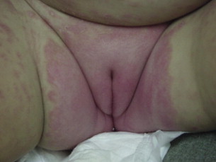
In contrast to contact dermatitis, psoriatic diaper dermatitis often presents with well-demarcated brightly erythematous plaques involving the diaper area, including the skin folds ( Fig. 2 ). Psoriasis involving the diaper area may respond poorly to low-dose topical corticosteroids used to treat common irritant diaper dermatitis. A family history of psoriasis can be helpful in making this diagnosis in infancy or early childhood. Keeping in mind the diaper area as a common location for psoriasis is also helpful when evaluating a confluent patch of scaling and erythema in this location.
In addition to diagnosing primary inflammatory dermatoses occurring in the diaper area, the possibility of infection should also be considered. Common infections may include candidiasis, bullous impetigo secondary to Staphylococcus aureus , and perianal streptococcal infection. Such infections should be included in the differential diagnosis of rashes in the diaper area, especially ones that do not respond to topical corticosteroids alone.
Infantile candidiasis should be suspected when a child presents with an erythematous patch or plaque (which may often be moist) involving folds in the diaper area; axillary skin can be involved as well. Patients may have surrounding scaling erythematous papules or pustules, often described as satellites. Potassium hydroxide smears or fungal culture should be diagnostic. In the healthy patient, topical azole antifungals are curative.
Staphylococcal diaper dermatitis usually presents as a superficial vesiculobullous process; often, patients present with well-circumscribed superficially eroded areas of skin, with minimal or no surrounding erythema ( Fig. 3 ). As with candidiasis in healthy infants, topical antibiotics can be useful. Staphylococcal scalded skin syndrome can be seen in patients of all ages. Tender and diffuse erythema is usually seen, with sparing of mucous membranes. As a result of the involvement of desmoglein 1, keratinized skin is the clinical target rather than mucous membranes in this toxin-mediated illness. The evolution is rapid, although, once the condition is diagnosed, appropriate antibiotic therapy results in prompt resolution.
Streptococcal disease can occur in the setting of group A beta-hemolytic streptococcal infections with typical facial erythema and circumoral pallor or as streptococcal diaper dermatitis. Characteristically, streptococcal diaper dermatitis is seen in a perianal distribution. Rather than purely epidermal erythema, psoriatic-appearing scale can be seen. Appropriate cultures and antibiotic therapy lead to (often) prompt resolution.
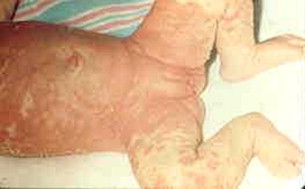
Rarer causes of diaper dermatitis that fail to respond to typical therapies include metabolic and nutritional deficiencies. Multiple nutritional deficiencies such as zinc deficiency, essential fatty acid deficiency, and malabsorption syndromes such as cystic fibrosis (CF) may also present with recalcitrant diaper, as well as more diffuse dermatitis ( Fig. 4 ). Zinc deficiency may occur secondary to acrodermatitis enteropathica, which is an autosomal-recessive genetic disorder that may occur sporadically because of nutritional zinc deficiencies. It typically presents with diarrhea, periorificial and acral dermatitis, and alopecia approximately 1 to 2 weeks after weaning from breast milk. It may progress to failure to thrive if not treated. The eruption and symptoms quickly resolve with zinc supplementation. Such patients classically present with a periorificial scaling dermatitis. Many have periungual involvement, with erythema and pustules. Pure zinc deficiency is usually associated with decreased levels of serum alkaline phosphatase because this enzyme requires zinc for its function. Essential fatty acid and biotin deficiencies can also resemble zinc deficiency clinically. Acrodermatitis enteropathica-like eruptions have also been reported in patients with malabsorption syndromes such as CF and in patients with other metabolic disorders such as organic acidurias. In CF, the same general pattern of erythema is seen. Clues to the diagnosis of CF in this setting can include larger than usual stools, often with mucus.
Testing for zinc and alkaline phosphatase, as well as serum amino and urine organic acids might be indicated, depending on the more likely diagnosis. Serum CO 2 is helpful to assess acid/base status, because many organic acidemias present with systemic acidosis.
Rarely, a persistent diaper dermatitis that does not respond to standard anticandidal therapies or topical steroids may be a sign of Langerhans cell histiocytosis (LCH). LCH is a rare disorder involving histiocyte infiltration of the skin plus possible infiltration of other organs, which can be fatal. Classically, it may present with purpuric macules or papules with hemorrhagic crusts in the diaper area, axillae, and scalp. Erosive areas on the gingivae can be seen in infants. The skin findings of LCH are most commonly misdiagnosed as seborrheic dermatitis or irritant diaper dermatitis in infants. Therefore, it is important to consider this diagnosis in diaper rashes that fail to improve after standard therapies. Extracutaneous manifestations such as lymphadenopathy, hepatomegaly, and bony lesions may be present. Diagnosis is based on a high index of suspicion leading to a biopsy that shows a Langerhans cell infiltrate with CD1a+ positivity on immunohistochemistry. A complete evaluation should include investigation of organ function and assessment of bones for possible involvement.
Localized dermatitis
Dermatitis may be localized to the diaper area or to periorificial areas in children, giving possible clues to the cause, including both common and rarer skin manifestations. Sometimes, the dermatitis may be more generalized. Seborrheic dermatitis may be more marked in skin folds and areas occluded by the diaper in infants. This condition is often confused with psoriasis. Whereas atopic dermatitis typically spares the diaper area, seborrheic dermatitis is commonly seen in the diaper area. Seborrheic dermatitis often presents in the first months of life with scaly red patches, often involving the flexural skin folds as well as the diaper area along with thick, greasy scale on the scalp. The condition can often present in a generalized fashion, in addition to the more common distribution ( Fig. 1 ). In contrast to atopic dermatitis, which is almost invariably pruritic, infantile seborrheic dermatitis is usually asymptomatic. Most cases of infantile seborrheic dermatitis clear easily with topical therapy such as low-potency topical corticosteroid. This condition should improve and resolve over the first several months of age.