Tendon transfers are performed predominantly to restore hand function or balance due to injuries of the radial, median, and ulnar nerves. Current surgical techniques for the most common tendon transfers for reconstruction of radial, median, and ulnar nerve palsies are demonstrated. These techniques can also be applied to restore flexion and extension of the fingers and thumb after injuries to the extrinsic flexor and extensor muscles and tendons of the forearm or intrinsic muscles of the hand.
Tendon transfers
Tendon transfers are reconstructive techniques that restore motion or balance to the hand that has impaired or absent function of the extrinsic or intrinsic muscle-tendon units of the forearm and hand. In a typical tendon transfer, the tendon of insertion of an expendable functioning muscle is detached, mobilized, and then reattached to another tendon or bone to substitute for the action of a nonfunctioning muscle-tendon unit. Occasionally, both the tendon of origin and the tendon of insertion are detached and then reattached at different locations. Unlike a tendon graft, the transferred donor tendon remains attached to its parent muscle. A tendon transfer also differs from a microsurgical free functional muscle transfer in that the neurovascular pedicle to the muscle of the transferred tendon remains intact.
There are 3 general indications for tendon transfers in the upper extremity:
- 1.
To restore function to a muscle paralyzed by injuries of the peripheral nerves, the brachial plexus, or the spinal cord
- 2.
To restore function following closed tendon ruptures or open injuries to the tendons or muscles
- 3.
To restore balance to a hand deformed by various neurologic diseases.
Tendon transfers are best conceptualized as a means to restore a lost function rather than a means of substituting for a specific muscle (ie, restoring strong pinch as opposed to restoring function of the flexor pollicis longus [FPL]). Tendon transfers are performed predominantly following peripheral nerve injuries and, therefore, the current techniques for reconstruction of radial, median, and ulnar nerve palsies are described. However, these same techniques can be used for posttraumatic reconstruction of the hand that is affected by injuries to the muscles and tendons of the forearm or hand.
General Principles
It is assumed that surgeons will already understand the general principles of tendon transfers, but these are briefly summarized:
- 1.
All fractures should be healed or rigidly fixed by internal fixation.
- 2.
All skin in the projected course of the transfer should be pliable and unscarred, otherwise it should be replaced by pedicle or free flap.
- 3.
Full passive range of motion of the metacarpophalangeal (MCP) and interphalangeal joints should be achieved by therapy or splinting before any tendon transfer.
- 4.
The muscle-tendon unit selected must be expandable so that the transfer does not create a new functional deficit. A minimum of 1 wrist extensor, 1 wrist flexor, and 1 extrinsic flexor and extensor tendon to each digit should always be retained.
- 5.
The potential amplitude or excursion of a donor muscle-tendon unit must be sufficient to restore the specific lost function :
- •
Wrist extensors and flexors: 33 mm
- •
Finger extensors: 50 mm
- •
Finger flexors: 70 mm
- •
The potential excursion of the donor muscle-tendon unit can be increased by 20 mm by the wrist tenodesis effect.
- •
- 6.
The tendon transfer should pass in a direct line from the origin of the donor muscle to its new insertion and should ideally only act across 1 joint.
- 7.
The tendon transfer should perform only 1 single function, but it may perform the same function in several adjacent digits.
- 8.
The donor muscle should be synergistic with the function of the muscle to be restored.
- 9.
The final selection of the potential tendon transfers is done by matching the available donor muscles with the functions to be restored.
Surgical Techniques
The success of any tendon transfer depends entirely on preventing scarring or adhesions along the course of the transferred tendon. Incisions should be carefully planned before elevation of the tourniquet so that the final tendon junctures lie beneath skin flaps rather than lying immediately beneath the incisions. The donor muscle should be carefully mobilized to prevent damage to its neurovascular bundle, which usually enters in the proximal third of the muscle. The transferred tendon should glide in a tunnel through the subcutaneous tissues and should not cross bare bone or pass through small fascial windows. Tendon junctures should be performed using a Pulvertaft weave technique. The donor and recipient tendons are sutured under appropriate tension, and after 1 or 2 nonabsorbable sutures have been inserted, the tension of the transfer should be checked by observing the flexion and extension of the digit during tenodesis of the wrist. Postoperatively, the hand is immobilized for 3 to 4 weeks, at which time gentle, active range-of-motion exercises are started, usually under the supervision of a therapist, but the hand is protected for a further 3 weeks in a light-weight plastic splint.
Timing of Tendon Transfers
Timing of tendon transfers may be classified as early, conventional, or late. A conventional tendon transfer is usually performed after reinnervation of the paralyzed muscle fails to occur by 3 months after the expected time of reinnervation, based on the rate of nerve regeneration of 1 mm per day. Brand, Omer, and Burkhalter have advocated early tendon transfers in certain circumstances so that a tendon transfer is performed simultaneously with the nerve repair or before the expected time of reinnervation of the paralyzed muscle. This early tendon transfer, therefore, serves as a temporary substitute for the paralyzed muscle until reinnervation occurs, by acting as an internal splint. If reinnervation is suboptimal, the early tendon transfer acts as a helper to augment the power of the partially paralyzed muscle, and if reinnervation fails to occur, it then acts as the permanent substitute.
Radial nerve palsy
Functional Deficits
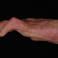
The functional motor deficit in radial nerve palsy consists of the inability to extend the wrist, the inability to extend the fingers at the MCP joints, and the inability to extend and radially abduct the thumb ( Fig. 1 A). However, another significant disability is that patients are unable to stabilize their wrist so that transmission of flexor power to their fingers is impaired resulting in marked weakness of grip strength (see Fig. 1 B). Unlike median and ulnar nerve palsies, sensory loss following radial nerve injury is not functionally disabling unless patients develop a painful neuroma.