Fig. 49.1
Pelvic computed tomography scan with oral contrast 7 days after low anterior resection. A collection containing contrast and air is seen posterior to the rectum
Another imaging modality, used less frequently today, is water-soluble contrast enema. The enema allows active visualization of the passage of contrast through the anastomosis and can determine if a disruption exists. If the anastomosis is located close to the dentate line, extra care should be given when placing the flexible catheter to prevent additional damage to the healing area. Alongside advanced imaging, these modalities also provide another therapeutic option to the clinician. If the patient does not present signs and symptoms of diffuse peritonitis and shock and a well-defined abscess is easily accessible, then percutaneous CT guided drainage is indicated [17]. This treatment is coupled with the additional placement of the patient on nutritional support and adequate antibiotic and antifungal coverage where the nonoperative approach can be successful and prevent a second major operation in selected cases.
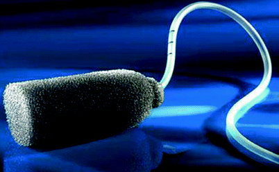
Recently, specialized vacuum-assisted drainage (the EndoSponge; B. Braun Aesculap AG, Germany) has been used with some success in localized leaks after a failed rectal anastomosis with a presacral collection [18, 19]. This usually results in rapid resolution of fever and bacteremia, with a mean duration of drainage of 21 days and an average of five sponge changes. Closure may take up to 2 months in many cases based on both radiological and endoscopic assessment, with the timing of closure dependent upon the age of the patient and the initial size of the cavity associated with the localized leak; larger cavities take longer to close. The more distal the anastomosis, the longer the closure time required. The place of a diverting loop ileostomy in such cases (if not previously performed) is unclear (Fig. 49.2).


Fig. 49.2
EndoSponge, a single-unit polyurethane sponge system used for selected cases of low rectal anastomotic leak with a presacral collection (Reprinted with permission from Arezzo et al. [19])
Overview of Reoperative Alternatives
After suspicion of a leaking anastomosis arises, it is the clinical experience and judgment that guide the course of action. In cases of diffuse peritonitis and systemic signs of sepsis, patients should be resuscitated and taken back to the operative room for exploration. If the clinical picture is unclear, imaging studies, as mentioned earlier, are indicated with the proper action to follow.
When a patient is taken back for exploration, the main operative strategy should include the following principles:
1.
Minimizing the extent of surgical intervention
2.
Shortening the procedure as much as feasibly possible
3.
Adequate abdominal washout
4.
Proximal fecal diversion should be favorably considered preoperatively with the relevant actions such as stoma markings
The surgical options and alternatives for a reoperative surgery are numerous but are guided by several factors. Generally speaking, the surgeon’s actions should be focused on saving the patient’s safety first with adequate quality of life second. Optional future interventions should also be taken under consideration but at a lower level of importance. The first step taken in most cases is reentering the abdominal cavity using the primary incision. Because of the peritonitis and the individual postoperative stage of the scar tissue, adhesions, and abscesses, cavities are encountered frequently. Extreme caution should be applied at this step to prevent additional damage. Slow and careful blunt dissection will induce fewer unnecessary injuries. Copious irrigation and maximal removal of enteric content follows, which will enable the identification of the “culprit” as well as minimizing the bacterial load and reducing the risk of abscess formation and septicemia.
Because of the friability of the tissues, diffuse peritonitis, and in some cases patient instability, repairing the anastomosis primarily cannot be considered or endorsed. Occasionally, primary repair seems feasible; however, this always has a poor healing prognosis because of widespread abdominal contamination. Hence, in most cases the shortest and safest procedure for anastomotic dehiscence will be an abdominal washout and proximal diversion. Primary repair is performed in some cases if the disrupted anastomosis is readily accessible, the colon appears relatively healthy, and the size of the defect does not indicate fashioning it anew. This is only considered in stable patients. Despite repairing the dehiscence, proximal diversion is still indicated to allow perfect conditions for the repaired anastomosis to heal.
Functional Outcomes of Redo Anastomoses
The functional reported outcomes of patients who have undergone a redo anastomosis after dehiscence are naturally limited. Many patients who undergo an emergent exploration and diversion never undergo definitive reconstructive surgery and remain with a permanent ostomy [20, 21]. The likelihood of a permanent stoma is greater after a colonic as opposed to a rectal leak, as is the attendant mortality [21]. In this respect, Lefevre et al. [22] recently have published their experience with redo surgery for failed colorectal or coloanal anastomoses. This study included patients with various reasons for secondary surgery—not all cases had a clinical leak. The authors reported a failure rate of 15 % (n = 5) of the newly fashioned anastomoses and concluded that although redo surgery is feasible, it is a demanding procedure with high short-term morbidity.
Special Situations
The Ileal Pouch
An ileal pouch anastomosis is performed for several indications when there is lack of colonic reserve, and its use places these patients in a special subgroup. Aside from the immediate risk to the patient, anastomotic leak in these cases has two significant long-term effects. Without the colorectal physiologic capabilities of water absorption and a reservoir, every segment of small bowel has extreme importance. A second operation and take down of the leaking pouch means a significant loss of small-bowel length and future difficulties in reconstructing a new pouch without compromising the quality of life. The other potential difficulty is the negative long-term effect of pelvic scarring and fibrosis. The result of the pelvic tissue reaction to the exposure to enteric content compromises the ability of the new pouch to stretch and expand, worsening the already compromised situation and compliance of the neoanastomosis. For these reasons, among others, most surgeons routinely will use a temporary diversion for patients undergoing an ileal pouch operation [23, 24]. The issue of reoperative surgery of ileal pouch anal anastomoses and the ability to save these pouches is a separate issue and is covered in Chap. 20.
The Colonic Pouch
The use of colonic J pouch (discussed elsewhere in this book) is intended to imitate the natural function of the rectum as a reservoir in the first 2 to 3 postoperative years. Unlike the ileal pouch operation, patients undergoing a colonic pouch usually still have a significant length of their colon, and in cases of a leak leading to a colostomy, an adequate quality of life can be maintained. Colonic pouches usually are covered by a proximal diversion and usually are salvageable with an ultimate ability to close the proximal stoma [25].
Irradiated Bowel
The topic of irradiated bowel is covered in Chap. 10


Stay updated, free articles. Join our Telegram channel

Full access? Get Clinical Tree


