Fig. 14.1
Breast retraction after breast conservation therapy (BCT) and radiotherapy without oncoplastic surgery of the inferior quadrant

Fig. 14.2
a Status after tumorectomy and radiotherapy of the left breast. The patient asked for bilateral remodeling. b Fibrosis and retraction of the left irradiated breast with a bad cosmetic result
14.3 Preoperative Planning
Radiological evaluation (mammography, breast ultrasonography, axillary ultrasonography, biopsy, MRI) of the breasts and distant metastasis research are major steps in the decision regarding which type of treatment to choose during the multidisciplinary counseling. MRI is the best imaging modality to evaluate the size of the tumor preoperatively and to find multifocal/multicentric lesions (which is a contraindication for BCT) and contralateral breast cancer [12, 13]. Thanks to these high-performance and preventive radiological evaluations, a greater number of small tumors are discovered, but 40–50 % of them are nonpalpable. These nonpalpable tumors are marked by ink or multiple hooked wires (harpoons).
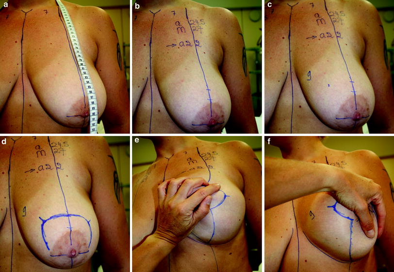
Preoperative photographs are taken in front view, three-quarter view right and left, and lateral view right and left. The drawings are done with the patient in a standing position. The midline from the sternal notch until the umbilicus is drawn. The tape measure is placed around the neck of the patient, and the axis of the breast is marked on the clavicle from the sternal notch, around 6.5–8.5 cm (for big patients) down to the abdomen (10–12 cm from the midline) (Fig. 14.3a). The new axis of the breast must not be influenced by the actual position of the NAC, which can be dislocated laterally or medially. The actual position of the NAC in centimeters is written on the patient. The sulcus is marked; it is an important landmark of the breast, and is always well marked during all the intervention as it is the limit of the inferior border of the new breast, the limit of our dissection. The new position of the nipple is marked horizontally on the axis of the breast by placing the index finger in the submammary fold. The superior border of the areola is 2 cm higher (around 18 cm in a short patient and up to 23 cm) (Fig. 14.3b). The position of the new areola is checked on the lateral view of the breast and must be located at the same level as the sulcus. The inner border of the new areola is marked 8–12 cm from the midline depending on the future breast volume desired by the patient (shorter distance for small breasts) (Fig. 14.3c). The outer border is drawn as a mirror image from the vertical axis of the breast (generally 4 cm but up to 5 cm in larger preoperative breasts). All these points are joined by a semicircular line of 8 cm to delimit the famous “mosquito dome” (Fig. 14.3d). The lower vertical limbs of the inferior skin resection are planned by pushing the breast outside and inside, very conservatively, and less than necessary in a breast reduction to avoid tension on the vertical scar and late dehiscence. The inner and outer vertical limbs are drawn as the prolongation of the axis of the breast previously marked on the abdomen (Fig. 14.3e, f). For the vertical scar alone, the two vertical limbs are joined 2 cm above the sulcus. For the inverted-T technique, 6 cm of the vertical limbs is conserved and the horizontal incision for the inverted T is drawn parallel to the inframammary folds. The inferior horizontal incision is made slightly above the sulcus with a triangle flap on the midline to avoid tension at the T. The breasts are examined apart and are pushed together on the midline to check the symmetry of the future inner incision. It is always very instructive to take a photograph with the design finished.


Fig. 14.3
Preoperative design of the superior pedicle technique with the vertical scar: a axis of the breast; b position of the new areola; c inner border of the new areola; d mosquito-dome drawing; e lateral vertical scar; f medial vertical scar
The patient is advised about the possible complications: the risk of immediate or later mastectomy when the margins are positive and if not enough breast is left in place for a cosmetic reconstruction.
14.4 Surgical Technique
The patient is placed in the supine position with the arms abducted for axillary access, with the possibility to seat the patient on the operative table to control the symmetry. General anesthesia is administrated and no local infiltration is used to prevent distortion of the operative site and tumorous cells spreading. Tumorectomy is performed with an incision/excision of skin in the skin excision of the breast reduction (Fig. 14.4


Stay updated, free articles. Join our Telegram channel

Full access? Get Clinical Tree


