CHAPTER 20 Superior and Medial Pedicle Breast Reduction Using a Vertical Pattern

Summary/Key Points
Introduction
The major advantage of vertical scar reduction mammaplasty is the improved long-term projection of the breasts following this procedure. With vertical scar reduction mammaplasty, the inferior wedge resection of the redundant breast tissue that contributed to breast ptosis and the subsequent suturing of the medial and lateral pillars, results in coning of the breast and a narrower, more projecting breast, which is the hallmark of this procedure.1
Lassus2,3 and Lejour4 are responsible for much of the pioneering work on vertical scar reduction mammaplasty. In 1969, Lassus2,3 developed a technique using a superior dermoglandular pedicle for transposition of the nipple–areola complex; a central en bloc excision of skin, fat, and gland; and a vertical scar to finish. The shape of the breast was produced by reapproximating the medial and lateral pillars with only suturing of the skin. In 1994, Lejour4 described a modification of Lassus’ technique in which liposuction was used pre-excision to eliminate fat contributing to breast volume; the skin surrounding the excised area was undermined; the superior dermoglandular pedicle was sutured to the pectoralis fascia; sutures were used in the breast parenchyma to reapproximate the pillars producing a more durable breast shape. Gathering of the skin of the vertical wound was used to keep the scar above the inframammary crease. In 1999, Hall-Findlay5,6 described a modification of Lejour’s technique using a mosque dome skin marking pattern; a full-thickness medial dermoglandular pedicle to transpose the nipple–areola complex; no skin undermining; no suturing of the pedicle to the pectoralis fascia; and liposuction only rarely to reduce breast volume. Hall-Findlay’s technique has since become the most commonly performed limited incision breast reduction technique as reflected in a 2002 American Society for Aesthetic Plastic Surgery survey of board-certified plastic surgeons.7
In 2006, we described our technique for vertical scar reduction mammaplasty that uses a mosque dome skin marking pattern; transposition of the nipple–areola complex on a superior or medial dermoglandular pedicle, depending on its position with respect to the skin markings; an en bloc excision of skin, fat, and gland; postexcision liposuction; and wound closure in two planes, including parenchymal pillar sutures and gathering of the skin of the vertical wound using four-point box stitches.1 Since 1989, we have performed this technique on over 2000 patients requiring breast reduction resulting in consistently good breast shape while leaving less scarring than more commonly performed breast reduction techniques.
Since Manchot’s8 1889 description of the blood supply to the breast, there have been many varying descriptions in the literature. Recently, van Deventer9 performed anatomical studies on 15 female cadavers in an attempt to further clarify the blood supply to the nipple–areola complex and found a large variation in the pattern of its blood supply. In all breasts, the nipple–areola complex received a blood supply medially or superiorly from one or more perforating arteries from the superior four perforating branches of the internal thoracic artery. The third perforator most frequently contributed blood supply to the nipple–areola complex in 47.5% of breasts, while the second perforator contributed in 25%, the first perforator in 15% and the fourth perforator in 12.5%. In 13 of 27 breasts, the nipple–areola complex did not receive any blood supply from superiorly. In 16 of 27 breasts, the nipple–areola complex received blood supply inferiorly from the fourth, fifth or sixth anterior intercostal arteries, with branches from the fourth anterior intercostal artery being the most common, occurring in 68.8% of breasts. The lateral thoracic artery contributed blood supply to the nipple–areola complex in 16 of 27 breasts, while the posterior intercostal arteries supplied the nipple–areola complex in only 1 of 27 breasts. In 2 of 27 breasts, the nipple–areola complex received blood supply from direct branches of the axillary artery. There were abundant anastomoses between these blood supplies around the nipple–areola complex. From this study, van Deventer et al10 concluded that the nipple–areola complex generally has a dual blood supply with the internal thoracic-anterior intercostal artery system providing blood supply from medioinferiorly and the lateral thoracic artery and other minor contributors providing blood supply from laterosuperiorly, with the most reliable source of blood supply arising from the internal thoracic artery.
Patient Selection
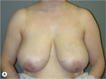
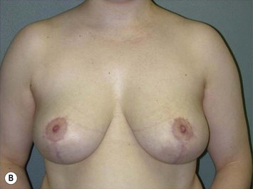
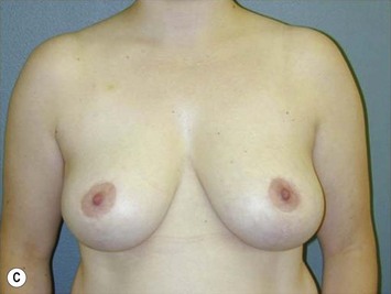
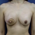
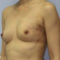
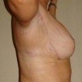
We have performed vertical scar reduction mammaplasty exclusively on over 2000 patients presenting for breast reduction (Fig. 20.1). In 2006, we performed a chart review of 250 consecutive patients treated between November of 2000 and December of 2003.1 In this clinical series, the average age of the patients was 38.5 years (range, 15 to 76 years) and the average body mass index was 28.8 kg/m2 (range, 17.3 to 46.3 kg/m2). The average weight of tissue excised per breast was 526 g (range, 10 to 2020 g). Liposuction was performed in 78.4% of cases and the average volume liposuctioned per breast was 140 ml (range, 50 to 500 ml). The average total reduction per breast (including liposuction when performed) was 636 g (range, 60 to 2020 g).
In addition, complications were analyzed based on body mass index, amount of reduction, pedicle selection, and use of liposuction.1 Although there was no statistically significant difference in the rate of complications between groups for amount of reduction, pedicle selection, and use of liposuction, there was a statistically significant difference between groups for body mass index with complications occurring less frequently in patients of normal weight (body mass index, 20.5 to 25.0 kg/m2). Since this study was published, we have limited performing this procedure to patients with a body mass index less than 35 kg/m2. Patients presenting for breast reduction with a body mass index greater than 35 kg/m2 are advised to lose weight prior to undergoing this procedure to decrease their risk for complications including superficial wound dehiscence and fat necrosis.
Along with other authors,3,5 we recommend learning this technique by initially operating on patients with mild to moderate hypertrophy and good skin quality. After becoming more familiar with the technique, one can progress to performing the technique on patients with more severe hypertrophy and poorer skin quality. Although it is possible to perform this technique on patients with extremely large breasts, it is important to realize that their postoperative breast size will likely remain larger when compared to other techniques because of the amount of skin preserved with a vertical scar reduction. In patients with severe mammary hypertrophy desiring a very small postoperative breast size, this technique is not suitable.
Indications
In an effort to establish clear, practical, objective, and fair criteria that could be applied by physicians to help differentiate women seeking breast reduction primarily for symptom relief versus aesthetic improvement, Kerrigan et al11


Stay updated, free articles. Join our Telegram channel

Full access? Get Clinical Tree


