Subcutaneous Pedicle Skin Flaps
J. N. BARRON
M. N. SAAD
EDITORIAL COMMENT
The Pers technique (Fig. 44.4) is a most elegant method using the nasolabial flap for reconstruction of nasal rim defects. It is much safer when the subcutaneous pedicle is based on a known arterial blood supply. The assurance of blood supply when performing the Pers technique for reconstruction of the alar rim is not as precise when compared to the use of a flap with a definite vessel.
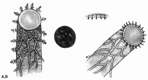
The subcutaneous pedicle skin flap consists of a skin paddle and a pedicle containing only subcutaneous tissue (1, 2, 3). Three donor sites are available for nasal reconstruction: (1) the nose itself, (2) the nasolabial area, and (3) the frontoglabellar region.
INDICATIONS
The nose, the nasolabial area, and the frontoglabellar region are all ideally suited for nasal reconstruction because they provide good color and texture matches. Although subcutaneous pedicle flaps are less robust than standard flaps, this is more than amply compensated for by their greater mobility and the small (but obvious) advantage of avoiding an unsightly dog-ear at the base of the flap. Also, scarring that results from the dissection of a subcutaneous pedicle is less than that which follows the use of a standard flap.
It is not advisable to repair very small defects with subcutaneous pedicle flaps, since there is the danger of mushrooming. In such instances, a graft or standard flap is preferable.
Nasal skin itself is particularly useful for the repair of lining defects. Because the nose has very little skin to spare, however, nasal skin defects alone are best reconstructed with flaps from the other two regions.
Subcutaneous pedicle flaps from the check adjacent to the nose can be used for the replacement of nasal lining, as well as for repair of skin defects of the lower part of the nose, especially the alae. Coverage of upper nasal, canthal, lower eyelid, and adjacent cheek defects is ideally performed with flaps from the frontoglabellar region. Although standard forehead flaps can be used, they have the disadvantages of limited mobility and the need for secondary correction of contour abnormalities at the base of the flap.
ANATOMY
It is not necessary to include an anatomically designated axial vascular system in the pedicle, although, if available, this is an advantage. Many subcutaneous pedicle flaps rely on a random capillary circulation, and because this is a low-pressure flow, it demands an ultra-atraumatic technique for dissection. We believe that the unilateral subcuticular suture disturbs the flap margin much less than ordinary interrupted sutures.
A direct vascular supply can be included with some subcutaneous pedicle skin flaps (4). Superiorly based nasolabial flaps derive their blood supply from the supraorbital and supratrochlear anastomoses, and the inferiorly based flap is nourished by the terminal branches of the facial artery or the superior labial vessels. Flaps from the frontoglabellar region can include blood supply from the supratrochlear vessels.
FLAP DESIGN AND DIMENSIONS
The function of the pedicle is to transmit blood, lymph, and nerve supply to the flap. Thus a good pedicle design is essential (Fig. 44.1). The pedicle should be long enough to allow the flap proper to reach the recipient site without the slightest tension.
The pedicle also should contain sufficient vascular elements to sustain the viability of the flap and, if possible, provide adequate sensation. The design should be such that kinking is avoided, and whenever possible, gravity should help venous and lymphatic drainage.
Stay updated, free articles. Join our Telegram channel

Full access? Get Clinical Tree


