Structure, Function, and Diagnostic Approach to Skin Disease


Structure, Function, and Diagnostic Approach to Skin Disease


Margaret A. Bobonich


Primary and nondermatology specialty care clinicians see the majority of patients with skin complaints on a daily basis. While patients make appointments to see their provider for a physical or blood pressure management, they commonly add an “Oh, by the way…” skin complaint. In contrast, many patients call the office or central scheduling and cite their reason for seeking treatment as a “rash.” This common, catch-all term reported by so many patients can leave the provider wondering what kind of eruption is really on the other side of the examination room door. Clinicians must be knowledgeable in evaluating skin lesions, which could range from skin cancer to a sexually transmitted infection.

Cutaneous lesions may represent more than just a skin disease and can be a manifestation of an underlying systemic process. Conversely, cutaneous conditions can cause systemic disease, dysfunction, and death. Psychosocial conditions can also be the cause or sequelae of skin conditions but are often negated. So in addition to maintaining competency in a primary specialty, clinicians need to acquire the essential knowledge and skills in dermatology—a daunting task given that there are almost 3,000 dermatology diagnoses.

The best approach for acquiring basic competency in the recognition and initial management of dermatologic disease (dermatoses) is to focus on conditions that are:


  • High volume—the most common skin conditions seen in clinical practice;


  • High morbidity—skin disease that is contagious or can impact quality of life or the community; and


  • High mortality—life-threatening conditions that require prompt recognition.

This chapter outlines essential dermatology concepts, including anatomy and physiology, morphology of skin lesions and algorithmic approach for the assessment of any skin condition. This will enable clinicians to develop the knowledge and decision making skills for far more than the 50 most common skin conditions. Subsequent chapters provide a comprehensive review of hundreds of skin conditions that MUST be considered in a differential diagnosis, ensuring an accurate diagnosis and optimal patient outcome.


STRUCTURE AND FUNCTION

Understanding the normal structure and function of the skin enhances your ability to correlate clinical and histologic findings associated with skin lesions (Figure 1-1). The skin is not only the largest organ but also the most visible, allowing both patients and clinicians the opportunity to observe changes and symptoms.

The skin is complex and dynamic and provides a physical barrier against the environment; an innate and adaptive immunity that protects the body from pathogens; and thermoregulation. The skin is also responsible for vitamin D synthesis and protection from ultraviolet radiation on non-hair-bearing skin. It is a reservoir for medication administration and is a sensory organ (pain, pressure, itch, temperature, touch). It comprises the epidermis, dermis, subcutaneous tissue, and adnexa or skin appendages and has regional variability in its thickness and structures. Glabrous skin does not have hair follicles or sebaceous glands, is located on the palms and soles, and is generally thick. In general, thin skin over the rest of the body houses a variable number of appendages, including the nails, hair, and sebaceous and sweat glands.


Epidermis

Commonly referred to as the “dead skin” layer, the epidermis is the locus of important structures and function (Figure 1-2). Cellular structures include keratinocytes, Langerhans cells, Merkel cells, and melanocytes. Nucleated keratinocytes differentiate as they ascend from the basal layer to the surface, filling with keratin and losing their nucleoproteins. Langerhans cells are intraepidermal macrophages responsible for phagocytosis of antigens and migration into the lymphatics and presentation to T cells. The immune function of the epidermis is paramount to our health. Merkel cells are believed to have a somatosensory function and are responsible for light touch and possible neuroendocrine function. Melanocytes synthesize the pigment which accounts for the variation in skin color among races. They are found in the dermis during fetal life and migrate to the basement membrane.

The layers (strata) of the epidermis are responsible for protecting the body from the environment as both a mechanical and chemical barrier. Each strata has unique characteristics and functions (Table 1-1). Flattened keratinocytes with a thickened cell membrane create the stratified layer (shingles on a roof) in the stratum corneum, which is not capable of metabolic activity. This cornified layer saturated in a lipid complex provides a virtually impermeable barrier and minimizes water loss. Thus, any defect or impaired function of this layer can lead to pathologic changes and disease.


Dermis

The dermis comprises fibroblasts, histiocytes, and mast cells, and is separated from the epidermis at the basement membrane (dermal-epidermal junction or DEJ). It adjoins with the papillary dermis (upper portion). Fibroblasts produce collagen (90% of the dermis), elastin, and ground substances, which comprise the majority of the dermis and are the supporting matrix of the skin (Figure 1-3). The dermis is also responsible for the continued immune response initiated in the epidermis by Langerhans cells, as well as neutrophils, lymphocytes, monocytes, and mast cells. Blood vessels, lymphatics, and sensory nerve endings for pain, itch, pressure, temperature, and touch are present. Arrector pili muscles in the dermis contract to make hair follicles stand up, creating the “goose bumps” effect. The reticular dermis (lower portion) joins with the subcutaneous or fat layer of the skin.


Subcutaneous Layer

The subcutaneous layer, also referred to as fatty tissue or hypodermis, comprises adipose cells and connective tissue, which varies in
thickness according to the body location. The hypodermis provides a layer of protection for the body, thermoregulation, storage for metabolic energy, and mobility of the skin.


FIG. 1-1. Skin anatomy and histology. A: Anatomy of the skin. B: Corresponding photomicrograph of the skin showing the cellular distinction between the epidermis and dermis.


Adnexa

Adnexa or appendages of the skin include the hair, nails, and eccrine and apocrine glands. The structure of hair and nails is discussed in chapter 14.


Glands


Eccrine glands

Chiefly responsible for thermoregulation of the body, the eccrine or sweat glands are tubules that extend from the epidermis through the dermis and are triggered by thermal and emotional stimuli. Although they are diffusely spread over the body, most are located on the palms and soles, and can contribute to hyperhidrosis or
hypohidrosis. Eccrine glands maintain an important electrolyte and moisture balance of the palms and soles.


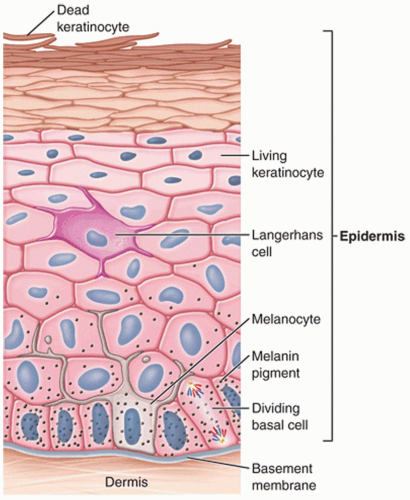
FIG. 1-2. Layers of the epidermis.
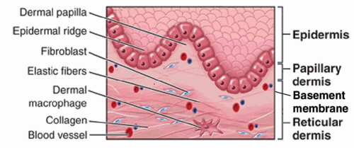


FIG. 1-3. Dermis.


TABLE 1-1 Strata of the Epidermis


STRATUM (LAYER)


CHARACTERISTIC


FUNCTION


Corneum


Brick and mortar layer


Lipid matrix and barrier


Antimicrobial peptides


Mechanical protection; limits transepidermal water loss; limits penetration of pathogens (bacterial, viral, and fungal) or allergens


Lucidium


Only on soles and palms


Protection


Granulosum


Keratin and fillagrin >80% of mass of epidermis


Profillagrin cleved into fillagrin, and loricin forming cornified envelop


Spinosum


Lamellar granules (containing ceramides)


Langerhans cells


Found intracellularly in upper layer but migrate to corneum where most effect, responsible for lipid barrier function


Defends against microbial pathogens


Basale


Cuboidal basal cells with nucleus and integrins


Scattered melanocytes


Integrins responsible for adhesion to dermis


Initiation of keratinocyte differentiation


Migration upward to stratum corneum takes 2-4 wk


Apocrine glands

Found only in the axillae, external auditory canal, eyelids, mons pubis, anogenital surface, and areola, apocrine glands secrete a minute amount of an oily substance that is odorless. The role of these glands is not clearly understood.


ASSESSMENT OF THE SKIN

Clinicians should elicit a good patient history and perform a proper skin examination in order to generate a complete differential diagnosis.

May 25, 2016 | Posted by in Dermatology | Comments Off on Structure, Function, and Diagnostic Approach to Skin Disease

Full access? Get Clinical Tree

Get Clinical Tree app for offline access