Steeple Skin-Muscle-Mucosal Flap for Lower Lip Reconstruction
M. F. STRANC
G. A. ROBERTSON
The repair of extensive lower lip defects has challenged the ingenuity of reconstructive surgeons for many years (1, 2, 3, 4, 5, 6, 7). The methods that have evolved can be broadly classified into three categories: those using lip tissues, those that rely on head and neck tissues, and those in which reconstructive material is brought in from a distance.
INDICATIONS
Satisfactory reconstruction of the lower lip must provide a lip curtain of adequate dimensions with sufficient muscular activity and sensory appreciation to allow normal speaking, drinking, and eating. It also must provide adequate access to all parts of the oral cavity and present an acceptable appearance.
Only locally available tissues are likely to meet these criteria. The problems become particularly acute when more than half the lip is missing. The steeple flap was designed to help this group of patients (8).
FLAP DESIGN AND DIMENSIONS
Following marking of the extent of the incision, the defect is converted into a rectangle, with the short side indicating the height of the lip resection and the long border marking the length of the excised lip. The island flap is marked out by extending the lower line of excision laterally for a distance equal to the height of the resected lip. Vertical sides of the island equal to the length of the resected lip are marked. A skin triangle is marked at the top to allow straight-line wound closure (Fig. 169.1A). The facial artery and its labial branches are located and marked. A Doppler device is of considerable assistance at this stage.
OPERATIVE TECHNIQUE
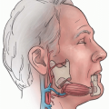
The lesion is excised as planned. The skin and subcutaneous tissues are incised around the flap. The incision is deepened to full thickness superiorly, inferiorly, and medially. The facial artery is identified on the superomedial aspect of the flap and tied. Tension on the tied vessel helps precise and safe location of the artery on the lateral side (Fig. 169.1B). Once the vessel is located, incisions above and below it are deepened to full thickness to within 5 mm of the vessel. Mucosa deep to the artery is divided, allowing transposition of the cheek tissues into their new site.
Related posts:


Stay updated, free articles. Join our Telegram channel

Full access? Get Clinical Tree


