Fig. 6.1
The 2-bladed square technique, (a) The purpose of the initial stage(s) is to define the lesion perimeter. The peripheral wound is sutured, (b) Tissue is processed to examine 100 % of the peripheral margins. After the peripheral margins are clear, the central island(s) is excised and the wound is reconstructed (Reproduced with permission from Arch Facial Plast Surg. 2001. 3(3):202–6. Copyright (2001) American Medical Association. All rights reserved [19])
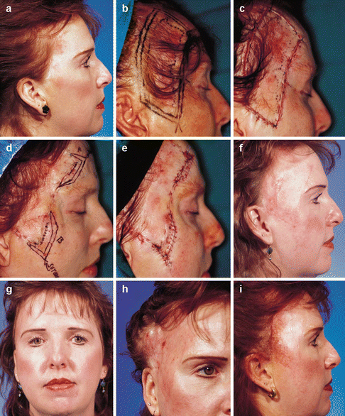
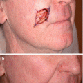
Fig. 6.2
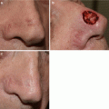
The 2-bladed square technique, (a) Locally recurrent amelanotic melanoma in situ located on the scalp, forehead, temple, and cheek of a 46-year-old patient who had been treated at least 7 times over 13 years. Treatments included multiple excisions (with slight atypical junctional melanocytic hyperplasia to the margins), cryotherapy, laser, and chemical peel, (b) Planned square excision with 1.0- to 1.5-cm margins. The central dotted line outlines the faint-pink lesion. The peripheral 2-lined rectangle outlines the peripheral lines of excision. A 2-bladed scalpel with 4.0-mm spacers is used for the procedure, (c) The tissue containing 100 % of the peripheral margin is excised and tagged with a suture for orientation. The specimen is paraffin embedded, and routine vertical sections containing 100 % of the peripheral margins are processed. The excision strip wound is sutured, (d) Two areas of positivity were identified (black dots). A thin strip of tissue containing 100 % of the peripheral margins was excised in a geometric fashion, with 1.0-cm margins around the areas of positivity, and again sent for processing, (e) The peripheral strip wound has been sutured. All peripheral margins were interpreted as negative for lesional atypical junctional melanocytic hyperplasia. The central islands of tissue were excised and sent to the pathology laboratory. The defect was repaired using bilateral supraclavicular full-thickness skin grafts, (f and g) Two-month postoperative result, (h) A tissue expander has been placed in preparation for scalp advancement to enhance the final cosmetic result and to recreate the natural hairline, (i) One-week postoperative removal of tissue expander and scalp advancement. No recurrence identified after 10 years (Reproduced with permission from Arch Facial Plast Surg. 2001. 3(3):202–6. Copyright (2001) American Medical Association. All rights reserved [19])
For the full square procedure, the entire lesion, including the 0.5 or 1 cm margin, is excised during the first stage. Once delivered to pathology, the peripheral margins are shaved again like a picture frame. All peripheral margins are processed and evaluated in the same manner as the 2-bladed square technique, while the center of the specimen is vertically serial sectioned. This method allows the pathologist to see the lesion from the center to the trailing edge, which may facilitate interpretation of the peripheral margin and help differentiate the malignant trailing edge from benign melanocytic up regulation often seen with chronic photodamage. This method may also be utilized for microstaging if significant clinical lesion remains, or in one stage combined with layered primary closure for lesions at lower suspicion for extensive subclinical extension.
The perimeter technique is similar to the 2-bladed square procedure [17]. Polygonal perimeter excisions and geometric staged excisions are variations of the full square procedure [16, 20]. The geometric staged excision differs from the full square only by use of a unique ink color at each peripheral margin specimen epidermal edge, and use of immunohistochemistry from the reported institution [20]. The use of immunohistochemistry with Melan A increased in the reporting institution over time from initially rare to standard for every case. No confirmed benefit of routine staining with immunohistochemistry of permanent sections exists. However, the dermatopathologists in this institution noted potential utility in assessment of melanocytic density at specimen margins in some cases. The need for immunohistochemistry for staged excision is the exception, not the rule.
Spaghetti Technique and Associated Variations
The “spaghetti” technique is almost identical to the 2-bladed square procedure [24]. It differs by the use of curved lines and rounded edges instead of straight lines and sharp-angled corners. Curved lines facilitate some tissue preservation (Figs. 6.3 and 6.4) [10, 13, 24, 28, 29]. Rounded edges may result in minimal loss of precision of margin associated anatomic location. The curved tissue specimens are pliable and can be pinned straight for perimeter en face sectioning, making the potential of false positive peripheral margins due to cutting deeper into the block with rounded instead of straight edges negligible.
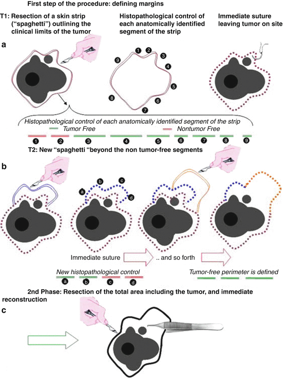
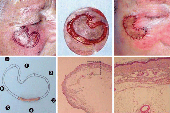
Fig. 6.3
“Spaghetti” technique, (a) A strip of skin along the surgical margin is resected, and is then divided into appropriately sized segments. The excised portion is sutured, leaving the central island intact, (b) Positive margins are identified and the process is repeated, (c) Once all margins are clear, the entire area, including the central island, is resected and the defect is reconstructed (Reprinted from J Am Acad Dermatol, Vol 64, Gaudy-Marqueste C, Perchenet AS, Tasei AM, Madjlessi N, Magalon G, Richard MA, et al., The “spaghetti technique”: an alternative to Mohs surgery or staged surgery for problematic lentiginous melanoma (lentigo maligna and acral lentiginous melanoma). p. 113–8. Copyright 2011, with permission from Elsevier [24])
Fig. 6.4
“Spaghetti” technique. Outlining limits of a lentiginous melanoma: Resection of the spaghetti, division into anatomically defined segments, suture of the defect (upper panel). Macroscopic appearance of the spaghetti segment together with histologic sections (lower panel) (Reprinted from J Am Acad Dermatol, Vol 64, Gaudy-Marqueste C, Perchenet AS, Tasei AM, Madjlessi N, Magalon G, Richard MA, et al., The “spaghetti technique”: an alternative to Mohs surgery or staged surgery for problematic lentiginous melanoma (lentigo maligna and acral lentiginous melanoma). p. 113–8. Copyright 2011, with permission from Elsevier [24])
A 2–3 mm wide strip of tissue is excised perpendicular to the skin like the outer ring of a dartboard, with the outer edge of the strip corresponding to the surgical margin. The central island containing the tumor is left intact and the marginal strip is sutured; the patient is left with no open wounds. The excised specimen is divided into appropriately sized segments, and a map is created for precise anatomic orientation. Each tissue specimen is stretched and pinned to convert a round specimen to a straight line specimen, processed with permanent vertical sections en-face for total peripheral margin control, and examined by a dermatopathologist. If areas of positivity are noted, the patient returns for subsequent procedures until all margins are free of disease. With each procedure a 2–3 mm wide strip of skin is excised around the positive margins, typically with up to 0.5 cm margins. After all peripheral margins are free of disease; the central island(s) is excised and processed with vertical serial sections. Definitive reconstruction is performed in this final stage.
Several variations have been described including a similar technique excising the entire lesion in the first stage identical to a full square, but with curved lines and rounded edges identical to the spaghetti method (Fig. 6.5) [13]. Another described this method with use of routine S100 and Melan A immunohistochemistry stains and digital pictures to facilitate orientation in cases with multiple stages [29].
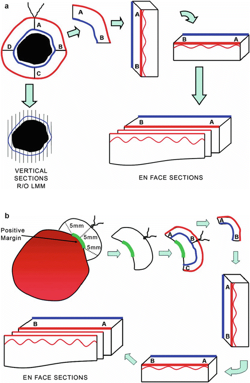
Fig. 6.5
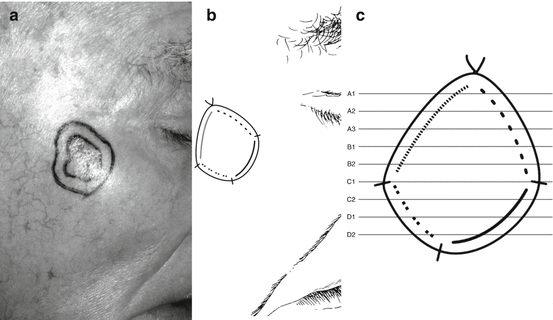
Variation of spaghetti and full square method with first stage excision of entire lesion with curved edges, (a) Representative diagram of the first stage of the staged excision procedure. The specimen is subjected to gross examination according to protocol and the margins are evaluated by en face sectioning, (b) Representative diagram of the second stage (re-excision) of the staged excision procedure. An additional 5-mm margin is taken from around the positive area, and the tissue is again subjected to gross examination according to protocol and the margins are evaluated by en face sectioning (Reprinted from Bosbous MW, Dzwierzynski WW, Neuburg M. Staged excision of lentigo maligna and lentigo maligna melanoma: a 10-year experience. Plast Reconstr Surg. 2009 Dec;124(6):1947–55. Reproduced with permission from Wolters Kluwer Health [13])
Slow Mohs
A modified staged surgery “slow Mohs” technique for LM treatment was first described in 1990 [30]. The technique as originally described involves excising the biopsy scar/tumor using the standard Mohs technique with a 45-degree inward bevel. The specimen is placed in formalin, divided, inked, mapped for orientation, processed for permanent sections cut horizontally in a standard Mohs fashion, and examined by a dermatopathologist. An added level of training, understanding, and expertise by the histotechnician is necessary to process the tissue with the deep and epidermal margin in the same horizontal plane. Additional expertise in interpretation of horizontal sections by the dermatopathologist is also required. One stage is performed per day with final reconstruction of the wound following clear margins.
Several variations have been reported since slow Mohs was first described [31–33]. One technique variation begins with the standard Mohs technique using frozen sections until all margins are interpreted as free of disease by the Mohs surgeon [31]. Another standard Mohs stage with a 1–3 mm margin is then performed and sent for permanent sections per slow Mohs above. The tissue is processed horizontally with the deep and epidermal margin within the same plane per the standard Mohs technique. If positive margins are noted by the dermatopathologist, another stage is performed excising tissue from the area of positivity with tissue processed with permanent sections as above. The process is repeated until all margins are interpreted free of disease resulting in a granulating wound, which may be repaired.
Another technique variation using permanent section margin processing for histologic evaluation was described [32]. Identical to the standard Mohs technique, a 45-degree inward bevel is used to excise the scar/tumor with a surgical margin. A 3–5 mm peripheral strip of tissue is dissected from the edges of the specimen and divided according to the map and corresponding skin scores placed during standard Mohs technique. The peripheral margin specimens are inked, mapped and processed in pathology for en face horizontal sections. The peripheral margin process is similar to spaghetti method previously described but with horizontal instead of vertical sectioning. The central specimen is serial sectioned vertically. If positive margins are noted, subsequent stages are performed until margins are free, at which time reconstruction of a granulating wound can be performed. Another similar technique using total circumferential margin control with both horizontal and vertical sections was reported [33].
Staged Excision with Radial Vertical Sections and Variations
A complex staged excision technique utilizing permanent vertical sectioning was described in 2008 [10]. The clinical lesion is excised using a standard vertical incision perpendicular to the skin surface. The peripheral margin is excised and divided into four quadrants and mapping is performed with orientation to the face of a clock (Fig. 6.6). A suture is placed at the 12, 3, 6 and 9 o’clock positions of each specimen, which is placed individually in corresponding formalin bottles. These 5 bottles are sent to pathology, along with a line drawing. The central debulk tissue is serially sectioned vertically at 2 mm intervals. The outer quadrants, the true surgical margin, are inked and vertically serially sectioned (not en face) at 2 mm intervals in a clockwise orientation. The permanent sections are evaluated by an experienced dermatopathologist. If positive margins are identified, or if tumor is noted within 2 mm of the peripheral margin in any section, a second stage with a 0.2–0.5 cm margin is obtained at 24 h. The specimen is processed as above with serial vertical sections and the process repeats until clear margins are obtained, at which point the patient returns and the granulating wound is reconstructed. A variation of this technique is reported where the central tumor is not debulked or separated from the true margin [12]. Instead, the lesion and the margin are excised en bloc, the specimen is mapped with orientation to the face of a clock. The specimen is placed in formalin and sent to pathology. The specimen is then bisected or divided into quadrants, which are then radially sectioned at 1 mm intervals [12].
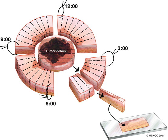
Fig. 6.6
Staged excision rush permanent section technique. Illustration shows tumor debulking and margins excised and evaluated with vertical sections with complete preservation of tissue orientation (Reprinted from McGuire LK, Disa JJ, Lee EH, Busam KJ, Nehal KS. Melanoma of the lentigo maligna subtype: diagnostic challenges and current treatment paradigms. Plast Reconstr Surg. 2012 Feb;129(2):288e–99e. Reproduced with permission from Wolters Kluwer Health [27])
Mapped Serial Excision
The mapped serial excision technique, first reported in 1998, simply involves more extensive serial sectioning (Fig. 6.7) [4, 11, 22, 23]. The specimen is excised in a typical fashion, tagged with a suture(s) for general orientation, mapped, inked, placed in formalin, and sent to pathology for permanent sectioning. The specimen is processed over 24 h with vertical bread loaf serial sections at 1–2 mm instead of standard 3–4 mm intervals. If positive margins are noted by the dermatopathologist, another excision typically with up to a 0.5 cm margin is performed at the area(s) of positivity with tissue processed again as above. The process continues daily until all margins are free of disease, at which time the granulating wound is repaired.
 Epidemiology and Natural History
 Incorporating Patient Preferences and Quality of Life
 Case B: Unsuspected Invasion and Upstaging in Lentigo Maligna Melanoma
 Emerging Novel Non-invasive Imaging
 Follow Up and Recurrence
 Case A: Multiple Mapping Techniques to Guide Staged Excision for a Challenging Lentigo Maligna Melanoma


Related posts:
Epidemiology and Natural History
Incorporating Patient Preferences and Quality of Life
Case B: Unsuspected Invasion and Upstaging in Lentigo Maligna Melanoma
Emerging Novel Non-invasive Imaging
Follow Up and Recurrence
Case A: Multiple Mapping Techniques to Guide Staged Excision for a Challenging Lentigo Maligna Melanoma
Stay updated, free articles. Join our Telegram channel
Full access? Get Clinical Tree
