Fig. 6.1Distribution patterns of atopic dermatitis in infancy.Courtesy, Julie V Schaffer, MD. From Bolognia JL, Schaffer JV, Duncan KO, Ko CJ. Dermatology Essentials, 1e. Philadelphia: Saunders, 2014, with permission.
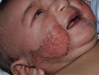
Fig. 6.2Atopic dermatitis, infancy. Acute lesions involving the lower cheek.Courtesy, Julie V Schaffer, MD. From Bolognia JL, Schaffer JV, Duncan KO, Ko CJ. Dermatology Essentials, 1e. Philadelphia: Saunders, 2014, with permission.
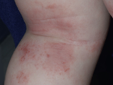
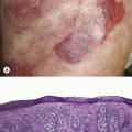
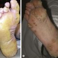
Fig. 6.3Atopic dermatitis, childhood. Acute to subacute lesions in the popliteal fossa.Courtesy, Julie V Schaffer, MD. From Bolognia JL, Schaffer JV, Duncan KO, Ko CJ. Dermatology Essentials, 1e. Philadelphia: Saunders, 2014, with permission.
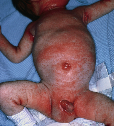
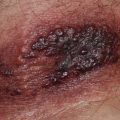
Fig. 6.4Infantile seborrheic dermatitis. Moist, ill-defined plaques favoring the body folds but also involving other sites in this case.Courtesy, Robert Hartman, MD. From Bolognia JL, Schaffer JV, Duncan KO, Ko CJ. Dermatology Essentials, 1e. Philadelphia: Saunders, 2014, with permission.
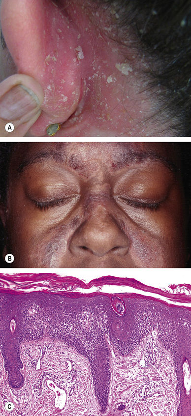
Seborrheic Dermatitis, Adult
Predilection for the scalp, posterior ears, central face, upper chest and back
Variably colored papules and plaques with flaking and/or greasy scale (Fig. 6.5)
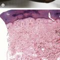
Fig. 6.5Seborrheic dermatitis, adult. A Erythema and flaky white scale behind the ear. B Purplish papules and plaques in a typical distribution over the central face. C Parakeratosis adjacent to follicles with intercellular edema (spongiosis).A, Courtesy, Norbert Reider, MD and Peter O Fritsch, MD; B, Courtesy, Jeffrey P Callen, MD. A,B, From Bolognia JL, Schaffer JV, Duncan KO, Ko CJ. Dermatology Essentials, 1e. Philadelphia: Saunders, 2014, with permission.
Only gold members can continue reading. Log In or Register to continue