The excellent cure rates associated with Mohs micrographic surgery depend on accurate interpretation of complete and high-quality microscopic frozen sections. Reliable interpretation of microscopic slides is only possible if the surgeon can distinguish tumor cells from surrounding normal tissue. By highlighting tumor cells with a chromogen that is visible on light microscopy, immunostaining allows the Mohs surgeon to distinguish tumor from normal cells in these challenging scenarios. This article focuses on practical aspects involving the most commonly used immunostains in dermatologic surgery, including MART-1 for melanocytic neoplasms, cytokeratin stains for keratinocytic neoplasms, and CD34 stains for dermatofibrosarcoma protuberans.
The excellent cure rates associated with Mohs micrographic surgery depend on accurate interpretation of high-quality microscopic frozen sections that allow examination of 100% of the microscopic margin. Reliable interpretation of microscopic slides is only possible if the surgeon can distinguish tumor cells from surrounding normal tissue. Although routine hematoxylin and eosin (H&E) frozen sections are usually sufficient, tumor cells can be difficult to detect on even the highest-quality H&E frozen sections if the cancer is poorly differentiated; if the tumor exhibits single cell spread; if tumor is surrounded by a dense inflammatory infiltrate; if tumor tracks along nerves, vessels, or fascial planes; if tumor is embedded in fibrotic tissue or connective tissue; or if there is pagetoid distribution of tumor. By highlighting tumor cells with a chromogen that is visible on light microscopy, immunostaining allows the Mohs surgeon to distinguish tumor from normal cells in these challenging scenarios. Several excellent comprehensive reviews of immunostaining in Mohs surgery have recently been published. This article focuses on practical aspects involving the most commonly used immunostains in dermatologic surgery, including MART-1 for melanocytic neoplasms, cytokeratin stains for keratinocytic neoplasms, and CD34 stains for dermatofibrosarcoma protuberans.
History of immunostaining in Mohs micrographic surgery
Immunostaining in Mohs surgery has evolved rapidly. In 1984, Robinson and Gottschalk published the first report of immunoperoxidase staining with cytokeratin antibodies on frozen sections during Mohs surgery for basal cell cancer (BCC) and squamous cell cancer (SCC). Although their immunostaining technique allowed improved identification of tumor cells with growth patterns that were challenging to detect on H&E slides, their complicated protocol required more than 24 hours to complete. Approximately one decade later, 2 groups independently reported successful treatment of BCCs and SCCs with anticytokeratin immunostaining protocols that required fewer than 90 minutes to complete. As immunostaining kits became commercially available and immunostaining protocols required fewer than 2 hours on frozen sections, surgeons expanded application of immunostains of Mohs frozen sections to other tumors, such as extramammary Paget disease, dermatofibrosarcoma protuberans, and melanoma.
Despite the proliferation of reports of immunostaining of Mohs frozen sections during the 1990s, only 13 of 108 laboratories run by members of the American College of Mohs Surgery reported the use of immunostains in a survey sent in 2000. The cost of reagents, the additional time required for both physicians and histotechnologists, and the additional expertise required for the application and interpretation of the stains may have deterred many Mohs surgeons from employing immunostains in their laboratories.
During the last 10 years, the number of reports of immunostains during Mohs surgery has proliferated, as documented by several excellent recent review articles. Bricca’s publication of a 1-hour protocol for immunostaining of melanoma frozen sections has inspired numerous investigators to innovate increasingly quicker protocols, including a 19-minute protocol for both cytokeratin and MART-1 immunostaining. As the time required to complete immunostains has decreased and immunostaining kits have become commercially available, Mohs surgeons can integrate immunostains into their practice with relative ease.
Controversies regarding the use of frozen versus permanent sections
Due to the wealth of data confirming superior cure rates when examining 100% of the microscopic margin, Mohs surgery with frozen sections has emerged as the gold standard compared with less thorough conventional methods of tissue processing in the treatment of BCC and SCC. While immunostaining may improve accuracy in the interpretation of frozen section microscopic margins, the percentage of BCCs and SCCs requiring immunostaining is relatively small. Consequently, scant data examine the efficacy of immunostaining in the treatment of these nonmelanoma skin cancers. Nevertheless, because the Mohs technique has proved so effective in the treatment of these tumors, little controversy arises with the addition of immunostaining to enhance the already excellent cure rates achieved with frozen section interpretation and H&E sections.
By contrast, the use of frozen sections to treat melanoma ignites considerable controversy. In comparison with BCCs and SCCs, immunostaining offers value and a practical advantage for the vast majority of melanomas. Distinguishing melanocytes from keratinocytes in the epidermis can be very challenging, especially if there is single cell proliferation and pagetoid spread of melanocytes or if there are surrounding atypical keratinocytes. Although melanocytes and keratinocytes have defining characteristics visible on H&E frozen sections, immunostaining offers more rapid and reliable identification of melanocytes at scanning magnification as compared with the H&E sections alone.
Perhaps because immunostains are a practical necessity for a higher percentage of melanocytic neoplasms, the volume of literature dedicated to frozen sections and immunostains for melanoma is much greater than for any other cutaneous neoplasm. Framing the controversies about frozen section immunostains for melanoma highlights several key points that apply to the treatment of many cutaneous neoplasms. First, many melanocytic tumors have microscopic extension that is not clinically visible. Second, if clinical examination is unreliable to determine tumor extent, then surgical margins based on clinical parameters are unreliable. Third, microscopic examination is the gold standard both to diagnose skin cancer and to determine margin status after excision. Fourth, the more thorough the examination of the microscopic margin, the more accurate the interpretation of margins status will be. Finally, microscopic examination is reliable only if tumor cells are clearly visible. Immunostains facilitate the identification of subtle tumor.
Substantial evidence demonstrates that clinical parameters are not sufficient in the diagnosis and treatment of melanoma. Because multiple benign pigmented lesions (eg, solar lentigines and atypical nevi) share clinical characteristics with melanoma and because a subset of melanomas defy clinical diagnostic criteria (such as the ABCDE criteria and the “ugly duckling” sign), diagnosis of melanoma is a complex process. In fact, the naked eye correctly diagnoses melanoma in only about 60% of cases. Another indication of the inaccuracy of naked eye examination is that dermatologists biopsy 18 benign nevi for every 1 melanoma. Microscopic examination is a practical necessity for the diagnosis of melanoma.
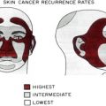
Surgical margins based on clinically visible parameters of melanoma are not reliable. Clinical and microscopic margins do not correlate in 90% of melanoma excisions. Sixty percent of the time, the true microscopic margin is less than the margin that the surgeon intended, based on clinical examination. Thirty percent of the time, the true microscopic margin is greater than the margin that the surgeon intended, based on clinical examination. Determining surgical margins is especially challenging on the head and neck, where the melanoma often arises in sun-damaged skin with multiple adjacent lentigines and keratinocytic growths. The high rates of positive margins and high recurrence rates after conventional surgical excision emphatically demonstrate that melanoma is a microscopic disease for which clinical parameters alone are insufficient to determine surgical margins. One out of 4 conventional wide local excisions on head and neck melanomas has positive microscopic margins, usually due to occult in situ melanoma. Recurrence rates after conventional excision of melanoma in situ on the head and neck range from 9% to 13%. The recurrence rates after conventional excision of lentigo maligna are even higher, ranging from 8% to 20%.
Compared with conventional excision, Mohs micrographic surgery offers superior local cure rates for melanoma. For melanoma in situ on the head and neck, multiple investigators have published local recurrence rates of only 0% to 2% after Mohs surgery. These superior local cure rates result because Mohs surgery allows examination of 100% of the microscopic margin, compared with examination of less than 1% of the microscopic margin with conventional methods of tissue processing. The consistently excellent local cure rates after Mohs surgery provide the greatest evidence of the accuracy of the technique. Nevertheless, opponents argue that frozen section examination of melanocytic lesions is inaccurate. Immunostains vastly improve one’s ability to detect melanoma, either with frozen or permanent sections.
It is important first to emphasize that Mohs surgery usually does not involve the diagnosis of the original tumor. Rather, the diagnosis is usually made via permanent sections prior to Mohs surgery. Therefore, the Mohs surgeon’s primary task is to determine whether tumor is present at the margin of the excision. The challenges of interpreting margin status for melanoma in situ apply to both frozen and permanent sections alike. When determining the presence or absence of melanoma in situ on H&E permanent sections, skilled dermatopathologists have only moderate agreement. Interpretation of melanoma margins poses similar challenges for both permanent and frozen sections.
Previous investigators have demonstrated that interpretation of melanoma margins using the highest-quality frozen sections is accurate in comparison with paraffin-embedded sections. Bienert and colleagues treated 97 patients with melanoma in situ or invasive melanoma of the face. In 25 patients, 117 tissue margins, which were defined as negative for melanoma at the time of H&E frozen sections, were reevaluated on H&E stains after formalin-fixation and paraffin-embedded tissue processing. There was 100% concordance between the negative status of the frozen and permanent sections in all 117 tissue margins. There were no cases of local recurrence in any of the 92 patients followed for a mean of 33 months. Similarly, Zitelli and colleagues demonstrated the reliability of using H&E-stained frozen sections to assess the margin status of 59 patients with lentigo maligna and melanoma. The investigators thawed 221 frozen sections from the 59 patients for paraffin sectioning. The H&E frozen sections had 100% sensitivity (ie, there were no cases where melanoma was detected on paraffin sections but not on frozen sections). There was 90% specificity of the H&E frozen section interpretation. In 4 patients, the specimens were read as melanoma or regressing melanoma by frozen sections, but paraffin sections showed only sun-induced epidermal atypia without melanoma.
Interpretation of margin status of melanoma by H&E sections alone is challenging, in part because melanoma in situ has some features in common with melanocytic hyperplasia in sun-damaged skin. Pagetoid spread, exocytosis of lymphocytes, and keratinocytic atypia make it especially difficult to distinguish between keratinocytes and melanocytes in the epidermis. Immunostains facilitate identification of melanocytes.
Immunostains in frozen sections allow detection of melanocytes as accurately as permanent section immunostains. Cherpelis and colleagues used a 19-minute MART-1 immunostaining protocol to compare melanoma-associated antigen recognized by T cells (MART-1) immunostained frozen and permanent sections. A board-certified dermatopathologist who was blinded to tissue preparation examined the slides. No significant difference was found in any of the following measurements: number of keratinocytes at dermal-epidermal junction, nuclear diameter of keratinocytes, numbers of melanocytes, melanocyte nucleus diameter, and melanocyte cytoplasm diameter. No significant difference was found in contiguity, pagetoid spread, melanocyte nesting, or atypical melanocytes. Because no statistically significant differences were observed in the measured variables between frozen sections and permanent sections, the investigators concluded that MART-1 immunostaining of frozen tissues is a reliable and useful technique with which to supplement Mohs micrographic surgery. Kelley and Starkus also found 100% correlation between frozen and paraffin-embedded sections of lentigo maligna stained with MART-1. Whether treating melanoma or other tumors, immunostains serve as a valuable adjunct to routine H&E-stained sections.
Related posts:
 Mohs Micrographic Surgery Technique
 Mohs Surgery for Squamous Cell Carcinoma
 Flaps and Grafts Reconstruction
 Management of Unusual Cutaneous Malignancies: Atypical Fibroxanthoma, Malignant Fibrous Histiocytoma, Sebaceous Carcinoma, Extramammary Paget Disease
 Special Considerations for Mohs Micrographic Surgery on the Eyelids, Lips, Genitalia, and Nail Unit
Multidisciplinary Approach to Large Cutaneous Tumors of the Head and Neck
Special Considerations for Mohs Micrographic Surgery on the Eyelids, Lips, Genitalia, and Nail Unit
Multidisciplinary Approach to Large Cutaneous Tumors of the Head and Neck
Stay updated, free articles. Join our Telegram channel

Full access? Get Clinical Tree


