Atopic dermatitis is the leading cause of pediatric dermatology visits in developed nations. Recurrent, itchy rashes in typical locations and a family/personal history of atopy helps to identify children with disease. Most cases (85%) are diagnosed by age 5 years. Some comorbidities are age-based and may affect disease course. Topical corticosteroids are the mainstay of therapy; corticosteroidphobia and side effects complicate use. Topical calcineurin inhibitors are alternatives to corticosteroids, especially in sensitive locations. Systemic therapies include antihistamines, immune suppressive agents, and phototherapy, with specific pediatric modifications. This article reviews the nuances and caveats of pediatric atopic dermatitis diagnosis and management.
Key points
- •
Atopic dermatitis (AD) is a chronic inflammatory skin and multisystem disease that affects children differently in different age categories.
- •
Consideration for the presence of comorbidities is important in caring for the pediatric AD patient.
- •
Infantile AD can be complicated by overlap with irritant contact dermatitis and seborrheic dermatitis.
- •
School-aged children with AD often suffer intercurrent infections with viral and bacterial pathogens.
- •
Teenagers with AD may have impaired body images and are more prone to specific types of allergic contact dermatitis.
Introduction
Atopic dermatitis (AD) is a multisystem inflammatory disorder that exists within the spectrum of diseases of atopy, that is AD, food and environmental allergies, and asthma, all of which are becoming more prevalent. Most atopic diseases begin in childhood, with 85% of AD cases starting by age 5 years and about one-quarter of children experiencing wheezing or eczema symptoms by their late teen years. The prevalence of atopic illnesses has increased 2- to 5-fold since the 1960s in developing countries in children and adolescents, with a recent estimate of 17.2% in 5- to 9-year-old children from Oregon. Mirroring this, asthma was noted to have a prevalence rising from the 1960s to the 1980s of 183 to 284 per 100,000, with the increase being accounted for by children ages 1 to 14 years, and especially increased for children with a parent who has had asthma. AD has a wide reaching effect on childhood and quality of life can be negatively impacted in pediatric AD, mirroring the severity noted with other pediatric chronic illnesses, such as renal disease and cystic fibrosis.
The increased prevalence of allergic illness has been accompanied by an increase in disease persistence, especially of severe AD, into the adult years. Factors associated with persistence are onset after 2 years of age and ongoing symptomatology for 10 or more years and females in metaanalysis ; therefore, it is crucial that we consider not just the youngest patients with AD, but the adolescent with long-standing disease, who may have ongoing symptoms for a lifetime.
This article is divided into practical categories in AD based on the age/developmental time period of the child, that is: (1) infancy, (2) toddler years, (3) preschool and school-aged children, and (4) preteens and adolescents. The focus is kept on the clinical nuances of the disease, and comorbidities expected for age and treatment considerations both in prescribing and side effect profiles, with an ultimate goal to improve care and, therefore, quality of life in pediatric patients with AD.
Infancy
Clinical Nuances
This brief overview includes common features noted in infancy that impact diagnosis and therapeutic considerations. The clinical nuances overlap with comorbidities, but are reviewed in only 1 section in the interest of space.
AD is defined as a pruritic eczematous condition with a chronic, relapsing course and a typical pattern of appearance, in infancy and early childhood of “facial, neck and extensor involvement,” and often accompanied by early age of onset, xerosis, and other forms of atopy. Eczematous plaques in infancy can occur anywhere, but are largely limited to the face and extensor extremities. Infants often scratch incessantly and before having the dexterity to scratch they will often rub or wiggle against surfaces to address itch. This action can be paired with significant sleep disturbance, especially in winter months when household heating decreases the relative humidity. Severity can range from a few limited plaques to erythrodermic appearance, which should be carefully differentiated from immunodeficiencies such as Leiner’s disease. Widespread disease in infancy is not uncommon owing to the impaired skin barrier and the thinner stratum corneum layer, allowing greater exposure to irritants and allergens. This phenomenon is reflected in clinical studies by an increased transepidermal water loss.
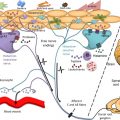
Although recent guidelines indicate that AD should be diagnosed in the exclusion of irritant contact dermatitis (ICD), allergic contact dermatitis (ACD), and seborrheic dermatitis (SD; Fig. 1 ), they do in fact overlap at times and these conditions may be more common in a child with AD or a predisposition to AD. AD is aggravated by skin contact with chemical and/or physical irritants such as excessive washing, soaps, and detergents. Facial AD in infancy ( Fig. 2 ) is generally complicated by an overlap with ICD caused by drool (aggravated with teething), messy eating, and the need for cleaning the face. Reduced indoor humidity can aggravate head and neck AD. Facial AD in infancy is usually associated with cheek eczematous plaques. The presence of lesions on the lower cheek, where the saliva might pool, or under a pacifier, may point to a larger component of ICD.
SD may overlap with AD in infancy and it has long been felt that the overlap is not random. Alexopoulos and colleagues have recently demonstrated that there is a true linkage of the 2 conditions. In their review of 87 children diagnosed with infantile SD (ISD) (mean age, 3.1 months), they were able to follow 49 children for 5 years, with 30 developing AD features at a later age—7 diagnosed with AD concurrent to ISD and 23 diagnosed with AD on average 6.4 months after ISD onset. The notable 3-fold increase in AD prevalence among ISD patients in this cohort highlights the fact that these 2 conditions cannot be separated and, as reviewed elsewhere in this article, therapy for AD, especially emollient-based care and irritant avoidance, should be initiated at ISD diagnosis. ISD in infancy often involves the skin folds as well, with maceration of the neck, and antecubital and popliteal areas, sites known to be affected by AD in older children. Yeast species including candida and Malassezia furfur can be isolated in these cases; therefore, the addition of an antifungal with candida coverage may benefit young children with AD and intertriginous disease.
There are a series of associations of infantile AD with environmental exposures that has been explored recently in the literature. Some of the environmental associations have included pesticide exposure, laminated wood floors, carpeting, urban environment, and home mold. Additionally, maternal food allergy and allergic disease, prenatal antibiotics, and prenatal stress may contribute to infantile AD occurrence. Ultimately, it seems that some activities such as nesting or redecorating may contribute to disease triggering in the susceptible infant.
Comorbidities
The original Hanifin and Rajka criteria included a list of almost 2 dozen minor features that have variable presence in childhood, depending on the age of the patient. A number of these minor criteria are comorbidities of disease. Those that are of significant concern in infancy include ISD and ICD (both discussed elsewhere in this article), food allergy, the Atopic March, and prurigo. Comorbidities of AD have recently been discussed in recent American Academy of Dermatology guidelines, which state that “Physicians should be aware of and assess for conditions associated with atopic dermatitis, such as rhinitis/rhinoconjunctivitis, asthma, food allergy, sleep disturbance, depression, and other neuropsychiatric conditions, and it is recommended that physicians discuss them with the patient as part of the treatment/management plan, when appropriate,” with a level of evidence of I, II (strength of recommendation C). An integrated, multidisciplinary approach was given level of evidence III with C strength of recommendation in the same guidelines; however, most children with severe AD do require integrated care.
Food allergy
A recent US-based study looking at the effect of early introduction of topical corticosteroids with or without pimecrolimus (initially blinded and then with open-label application) as a primary prevention of the atopic march in children 3 to 18 months of age with mild to moderate AD looked at the incidence of food allergies in this population. By the end of study, 15.9% had developed at least 1 food allergen, namely, peanut (6.6%), cow’s milk (4.3%), and egg white (3.9%); seafood, soybean, and wheat allergies were rare. A single recent study (the LEAP trial [Induction of Tolerance Through Early Introduction of Peanut in High-Risk Children]) has identified a 4- to 5-fold reduction in peanut protein allergy since age 5 years with early peanut protein; however, allergen screen and introduction are best conducted with an allergist in the setting of known or suspected peanut allergy. Avoidance of known food allergens in skin care products is advised as well in the setting of situations where there is an unknown refinement process and the presence or absence of the allergenic component is unknown. Food allergy testing is generally performed in infants with AD when there is (1) severe disease and/or persistent disease poorly responsive to topical care or (2) known food triggers. We further recommend allergy testing in the setting of potentially problematic nutrition issues owing to restrictive diets. Restrictive diets have not been recommended in the management of moderate to severe AD in the absence of proven allergens.
The atopic march
The atopic march refers to the theory that AD in childhood and the associated abnormal skin barrier may allow for the development of food allergy and asthma, that is, a march from the skin to other forms of atopy. The theory has some supporting evidence, although it may not describe or occur in all patients.
A variety of studies have looked at the atopic march in childhood from alternative perspectives. For example, a recent study supports the idea that percutaneous sensitization to foods may play a role in AD children, owing to an impaired skin barrier. Household dust has been identified as a potential source of peanut protein sensitization in children ages 3 to 15 months. The exposure–response relationship between peanut protein levels in household dust and peanut skin prick test sensitization is noted especially in children with a history of AD (odds ratio, 1.97; 95% confidence interval, 1.26–3.09; P <.01) and severe AD (odds ratio, 2.41; 95% confidence interval, 1.30–4.47; P <.01).
The Stop Atopic March trial, a 6-year study that addressed the concept of tight AD skin control in infants as a means of allergy prevention, compared 3- to 18-month-old infants who for 3 years enrolled in a double-blind study in which they were randomized to pimecrolimus or vehicle and later received 3 years of open-label pimecrolimus. The observed mean was 2.8 years and there were no differences between the groups, with 37% developing comorbidities: asthma (10.7%), allergic rhinitis (22.4%), food allergy (15.9%), and allergic conjunctivitis (14.1%). Because both study arms offered fair disease control and barrier repair, it may be that the study design was not adequate to reveal potential therapeutic interventions needed to truly stop the atopic march.
Topical and Oral Medication Risks and Benefits
Topical emollients and gentle skin care
The abnormal skin barrier in infants with AD is of particular concern owing to the intrinsically thinner skin and great irritation risk within the infantile age group, noted largely owing to incomplete barrier development at birth. One of the most elucidating studies of the past few years has been a clinical trial offering at risk infants (1 parent with AD) emollient versus no emollient daily from early infancy (by 3 weeks of age) and onward. At 6 months, the intervention resulted in approximately 50% reduction in AD. Furthermore, many groups have highlighted the need to avoid fragrance in at risk or AD infants. Recent guidelines from the American Academy of Pediatrics subsection on dermatology have recommended every 2 to 3 days bathing for 10 to 15 minutes, lukewarm water, gentle cleanser (fragrance-free Syndet or moisturizer enhanced), and daily emollient use. This is the cornerstone of therapy throughout life. Additionally fragrance-free, dye-free detergents, humidification for indoor heated homes, and cooling the home in hot summer months may enhance care.
The eczema action plan
The eczema action plan is a goal-directed direction sheet that helps parents to recount the discussion of the office visit regarding skin care and preventive measures as well as to empower parents to initiate treatments at the first sign of flare. There are approximately 10 parameters that may be included in these documents: (1) cleansing techniques and choice of cleansers, (2) application of standard topical agents, one for the face/groin and one for the body, (3) rescue medication (a stronger topical therapy for resistant areas), (4) emollient application, (5) oral antihistamines use, (6) use of topical antibacterial agents including mupirocin and bleach bathes, (7) detergent choice and clothing/fabric type, (8) control of temperature and humidity, (9) oral antibiotics when needed, and (10) other considerations, including sunscreens, allergen avoidance, and avoiding individuals with cold sores. A variety of resources exist online that demonstrate potential eczema action plans ranging from flare reduction plans to global skin care plans, all of which may be used by practitioners to enhance communication and compliance.
Topical medications
The topical therapy of infantile AD is largely management of the disease using the lowest potency topical corticosteroid to reduce and eliminate the localized disease flare. This generally means the use of class 5 or 6 topical corticosteroids for the face and intertriginous areas and class 3 or 4 topical corticosteroids for nonfacial areas. Rescue medication with a class 2 corticosteroid may be used occasionally, especially for severe flares of disease.
Risks in infancy include inadequate clearance, induction of parental corticosteroid phobia, and less commonly the true side effects of topical corticosteroids, including absorption and hypothalamic–pituitary–adrenal axis suppression, growth suppression, atrophy, and cataracts. Topical calcineurin inhibitors that are labeled as not intended for use under the age of 2 years have been endorsed as having evidence in support of their use for children under the age of 2 years who do not respond to topical corticosteroids. There are now some published data on the safety of pimecrolimus 1% cream in particular for children with AD for 5 years, that supports infantile use through early childhood.
The black box warning on the medications indicates a theoretic risk of skin cancer and lymphomas with topical calcineurin inhibitors, which may concern some parents greatly and prevent their use accordingly.
Topical mupirocin 2% ointment is not a primary therapy for AD, but can be used in children with concomitant bacterial superinfection of the skin and AD and has been paired with oral antibiotics and activities such as bleach baths to prevent recurrent bacterial superinfection in AD.
Oral medications
Oral medications have a place in all age groups of pediatric AD. On occasion, oral cyclosporine is used; however, the vast majority of children in the infantile age group are managed with topical agents and concomitant allergy screening/intervention. Oral antihistamines are used to promote somnolence in children with severe pruritus in association with AD. Sedating antihistamines include diphenhydramine and hydroxyzine. Dosage increases owing to relative reduction in efficacy should be avoided and avoidance in asthmatics with active symptoms owing to the risk of respiratory suppression is recommended. Paradoxic hyperreactivity is most frequent in younger children and signals the need to avoid the class of sedating antihistamines.
Oral antibiotics should only be used in the setting of extensive skin weeping, oozing, and/or pus discharge paired with a positive culture (usually Staphylococcus or Streptococcus ). In this setting, antibiotics such as cephalexin or oxacillin that have dual Staphylococcus or Streptococcus coverage are desirable, and cultures should be performed to identify potential need for therapy of methicillin-resistant Staphylococcus aureus .
Toddlers
Clinical Nuances
AD clinical manifestations varies with age. Although dermatitis involving the face, trunk, and/or extensor extremities predominates in infants, flexural surfaces like the wrists/ankles and antecubital/popliteal fossae are more common in toddlers and preschool- and school-aged children. It may also present with other features. The knowledge of the prevalence of less common clinical manifestations of AD according to age in different populations might be helpful in diagnosing incipient cases of AD.
Dennie–Morgan fold
Toddlers and preschool children may present with a Dennie–Morgan fold, a gross infraorbital fold caused by repeated scratching and rubbing of the face. It is considered by Hanifin and Rajka a minor criteria and should suggest atopy at first sight. Although this finding can be linked to allergic conjunctivitis, it is in fact noted more commonly in children of color, irrespective of the presence of AD and/or allergic conjunctivitis.
Diaper dermatitis
In infants, the “diaper area” ( Fig. 3 ) is often triggered by frequent cycles of skin wetting and drying as well as exposure to endogenous (eg, drool, urine, and feces) or exogenous irritants (eg, cleansing products or components of the elastic border of diapers). The latter are more common in toddlers with ACD.
Comorbidities
Irritant contact dermatitis
In pediatric patients, ICD is most common on the face, dorsal aspect of the hands, and “diaper area,” often triggered by frequent cycles of skin wetting and drying. The most effective way to alleviate ICD is with strict avoidance of likely triggers. When triggers cannot be identified or avoided, or there is residual dermatitis after triggers have been removed, mild topical corticosteroids may reduce inflammation.
Eczema Coxsackium
Eczema Coxsackium was reported in 2013, attributable to Coxsackievirus A6, and is being recognized increasingly. In contrast with the oral erosions and gray–white, oval vesicles on the hands, feet, and buttocks typically associated with hand–foot–mouth disease, eczema Coxsackium manifests as hemorrhagic vesicles within dermatitic skin. Children with AD typically have associated fever and constitutional symptoms as well as subsequent onychomadesis. A diagnosis of eczema Coxsackium is confirmed by serum polymerase chain reaction for Coxsackievirus. Treatment is supportive, with antipyretics and bland skin care.
Perianal bacterial dermatitis
Perianal streptococcal dermatitis, predominantly affecting children and particularly younger children, is most commonly caused by group A beta-hemolytic streptococci. The clinical picture of a sharply demarcated perianal erythema is very characteristic. The diagnosis is made by either a swab of the affected region submitted for microbiological analysis with the specific question for group A beta-hemolytic streptococci, or a rapid strep test. Systemic antibiotics such as penicillin, erythromycin, newer macrolides, or others, probably augmented by topical antiseptic or antibiotic ointments, are the treatment of choice. Treatment duration should be at least 14 days or, even better, 21 days, and be dictated by clinical and microbiological cure. Recently in the United States there has been a notable increase in perineal and perianal S aureus infection with dermatitis, often associated with AD. These cases sometimes have mixed Staphylococcus and Streptococcus overgrowth and present with less sharply demarcated lesions and more pruritus and eczematous changes. Culture becomes important; however, milder cases of the latter can be treated like superinfected AD.
Topical and Oral Medication Risks and Benefits
Nuances in topical therapeutics for toddlers and school-aged children are similar to toddlers, with the exception of the cases as noted. Specific review of therapeutics for unusual presentations was reviewed elsewhere in this article.
Preschool and school-aged children
Clinical Nuances
Pityriasis alba
Pityriasis alba is a common, idiopathic asymptomatic condition. Pityriasis alba is associated with AD as a minor diagnostic criterion. Pityriasis alba is most often present in school-aged children with no gender bias. Poorly circumscribed, hypopigmented patches are most often on the face and proximal upper extremities. Patches become more prominent during summer sun exposure as the surrounding skin tans, and it is more apparent on darker skin tones. In winter, hypopigmentation is less prominent. Emollients can minimize scaling, but will not impact hypopigmentation, which may persist for months despite control of xerosis. A few reports have documented efficacy of pimecrolimus cream 1%
Nummular dermatitis
Nummular dermatitis is characterized by well-demarcated round or oval plaques (ie, nummular), usually asymmetrically distributed on the limbs ( Fig. 4 ). Unlike typical AD, nummular dermatitis is unusual before age 5 and itch is not a common symptom. The cause of nummular dermatitis is unknown, but because many cases are associated with microtrauma (eg, insect bite, scratching), and other suspected factors which include S aureus colonization, contact allergens, or irritants and xerosis.