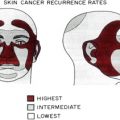
Although skin cancers on the eyelids, lips, genitalia, and the nail unit are infrequent, some skin cancers can have higher recurrence rates in these anatomic locations. Intrinsic to Mohs micrographic surgery (MMS) is maximal tissue preservation and high cure rates, making it a worthwhile procedure for these sensitive anatomic areas to ameliorate any functional or aesthetic compromise. The Mohs surgeon can provide a great service to patients by being aware of the anatomy, specialized instruments, unique histology, and potential complications when performing MMS on these areas.
The American Academy of Dermatology’s Guidelines of Care for Mohs Micrographic Surgery were developed to promote the continued delivery of quality care to our patients by using the Mohs technique for certain tumors and on specific anatomic sites where complete removal of the tumor with maximal normal tissue preservation and the lowest recurrence risk is needed. The need to maximize normal tissue preservation on the eyelids, lips, genitalia, and nail unit apparatus is of the utmost importance for obvious reasons, including the need to maintain free margins, functional competence, and aesthetic values. Basal cell carcinomas (BCCs) may have a potentially higher risk of recurrence with other types of treatment modalities in the periorbital and perioral areas. Likewise, squamous cell carcinomas (SCCs) of the periorbital skin/canthus, genitalia, lip, and nail bed/matrix have a higher risk of local recurrence. Less common tumors that favor these areas, such as verrucous carcinoma, sebaceous carcinoma, microcystic adnexal carcinoma (MAC), extramammary Paget disease, and erythroplasia of Queyrat, may also be successfully treated with Mohs micrographic surgery (MMS).
When the choice is made to proceed with MMS for tumors at these sites, there are other considerations that the prudent dermatologic surgeon must take into account to have optimal outcomes. The unique anatomy, free margins, and specially adapted form and function at these sites must be appreciated to remove the tumor and minimize collateral damage and thus lessen any potential side effects for the patient. Often, specialized instruments may be needed to work on these locations. These sites may contain transitions between thicker, hair-bearing skin and thin glabrous skin, with more laxity, which can sometimes challenge the inexperienced surgeon to remove the layers without damage or scalpel chatter, and at an optimally consistent level. Because of the lower prevalence of skin cancers at these sites (ie, MMS may not be performed so frequently in these anatomic locations), the occurrence of rare tumors, and given the unique histology of mucosal skin and nail matrix, the pathology on frozen sections, can be more difficult to interpret. Transitions from acral skin to nail matrix, mucosal eyelid to tarsal plate to cutaneous eyelid skin, or cutaneous lip to mucosal lip can frequently require more care when positioning, embedding, and sectioning tissue specimens, so experienced Mohs histotechnicians should be used whenever possible. Because of the superficiality of skeletal muscle and generally excellent vascular supply to these areas, bleeding is always a concern when performing surgery at these sites. In particular, electrocautery/coagulation for hemostasis must be used carefully and judiciously near the cornea and nail matrix to prevent accidental scarring to the eye or permanent undue dystrophy of the future nail plate.
The remainder of this discussion details site-specific tumors amenable to MMS and also briefly mentions pertinent anatomy, site-specific instruments, potential adverse outcomes, and repair options for these locations. Many books and journal articles cover these subjects in more depth.
Eyelids/periorbital
The eyelids are composed of cutaneous skin (<1 mm thick), orbicularis oculi muscle, the tarsal plate, and the conjunctiva. At the eyelid margin the skin tightly adheres to the tarsal plate. In the vertical dimension the inferior tarsal plate is 3 to 5 mm tall and the superior tarsal plate is 10 to 12 mm high. The tarsal plates connect to the bony orbital rim by the medial and lateral canthal ligaments. These ligaments can be cut intentionally for more laxity if needed (eg, for a large wedge repair), or sutured to different areas of the orbital rim to perform a canthopexy. The lacrimal gland with its canaliculi sits in the superior and lateral area of the upper eyelid, and the lacrimal caruncle, canaliculi, and sac are located in the medial/inferior canthus area. If these features are inadvertently damaged during surgery they can cause dry or watery eyes, respectively.
Specialized tools used for MMS on the eyelids include, but are not limited to, corneal shields (we favor plastic shields over the metal ones used for laser to eliminate the risk of inadvertent damage to the cornea and sclera during electrocoagulation), chalazion clamps, small curved iris scissors, Castroviejo scissors and needle holder, Bishop-Harmon forceps, Stevens tenotomy or gradle scissors, micro/beaver blades, 15-c scalpel blades, and micro-tipped/fine-tipped, Teflon-coated, electrosurgery tips.
Approximately 5% to 10% of skin cancers occur in the periocular region ( Table 1 ). Risk factors for periocular neoplasms include advancing age, fair skin, family history, and chronic sun exposure. The most common malignancies are BCC (90%), sebaceous carcinoma (5%), SCC (4%), followed by malignant melanoma (1%), lymphoma, Kaposi sarcoma, MAC, and rarely Merkel cell carcinoma ( Box 1 ). BCCs (∼70% of the time), SCCs (∼68%), and melanoma (∼57%) occur more commonly on the lower lid. On the other hand, sebaceous carcinoma occurs on the upper eyelid 63% of the time because the upper eyelid has more sebaceous glands than the lower lid. Most BCCs and SCCs involve the lower eyelid, followed by the medial canthus or upper eyelid and then lateral canthus. Eyelid BCCs, SCCs, sebaceous carcinoma, and Merkel cell carcinoma have all been treated with traditional frozen section MMS, and malignant melanoma has been treated with modified slow Mohs with paraffin-embedded tissue, all with lower recurrence rates versus surgical excision ( Table 2 ).
| Periorbital/eyelids | 5–10 |
| Lips | 3–4 |
| Penis | 0.5 (of all cancers) |
| Vulva | 1 (of all cancers in women) |
| Nail unit | ∼0.3 |
Periocular/eyelids
BCC
SCC
Sebaceous carcinoma
Melanoma in situ
MAC
Merkel cell carcinoma
Lips
BCC
SCC
SCC in situ
MAC
Genitalia
SCC
BCC
Extramammary Paget disease
Dermatofibrosarcoma protuberans
Granular cell tumor
Nail unit
SCC
Keratoacanthoma
Melanoma in situ
| Site | Tumor | MMS (%) | Median or Mean Follow-up (Years) | WLE (%) | Median or Mean Follow-up (Years) | References |
|---|---|---|---|---|---|---|
| Periorbital | SCC | 0–3.6 | 2.75–5 | 6+ | 2.6–4 | |
| Periorbital | BCC | 0–2 | 5 | 5–26 | 2.6–4 | |
| Periorbital | Melanoma in situ | 0–5 | 2–5 | 21–25 | 2–5 | |
| Periorbital | Sebaceous carcinoma | 11–12 | 3.1 | 9–36 | 5 | |
| Periorbital | MAC | 5–12 | 5 | 40–60 | Variable | |
| Lip | SCC | 0–8 | 5 | 10+ | Variable | |
| Lip | BCC | 0–3 | 3–5 | 12.5 | 2.6 | |
| Genitalia | Extramammary Paget disease | 16–23 | 3.25–5 | 31–61 | Variable | |
| Penis | SCC | 5.9–32 | Variable | 3–30 | Variable | |
| Vulva | SCC | 27 | Variable | 38 | 2.6 | |
| Nail unit | SCC | 4–8 | 5 | 5 (WLE) 56 (LSE) | <3 | |
| Nail unit | Melanoma | 21 | 7.7 | 0–11 (with amputation) | Variable |
Adverse outcomes that may occur whenever surgery is attempted on the eyelids include the normal complications seen in any skin surgery repair such as scarring, dehiscence, flap failure or graft necrosis, hematoma, and infection. Site-specific complications may include ectropion, trichiasis, lagophthalmos, ptosis, keratoconjunctivitis, sicca, watery eye, webbing, entropion, and orbital hemorrhage.
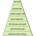
Repair options for Mohs defects on the eyelids or periocular skin include, but are not limited to, secondary intention healing, direct closure, grafting, pentagonal excision with direct closure, lateral canthotomy and inferior cantholysis with direct closure, Tenzel semicircular advancement flap, Mustardé laterally based cheek rotation flap, Hughes tarsoconjunctival flap, hinge flap with or without cheek advancement flap, Cutler-Beard bridge flap, glabellar flap, forehead flap, and the spiral flap.
It is important to consult or refer to oculoplastic colleagues when a repair is beyond the skill or expertise of the Mohs surgeon. If the Mohs surgeon is comfortable performing the repair, it is important to pay close attention to suturing technique, placing any sutures or knots on the cutaneous side of the eyelid, including throwing the deep sutures with the knots facing outward and away from the cornea so as to not cause abrasion and resultant scar. This technique is counter to the typical dermal buried suture technique. Here, suture is passed through the wound edge superficial to deep and deep to superficial so that the knot is more superficial and not buried toward the cornea. It is also important to close the muscle layer and cutaneous layers, paying attention to tarsal plate alignment. The mucosal side can be closed via a buried or subcuticular suture; however, if the muscle and cutaneous eyelid are well approximated, the mucosal eyelid often heals nicely by secondary intent. If necessary, a Frost suture (a horizontal mattress through the lower lid taped to the superior brow) may be used to reduce downward traction and possible cicatricial ectropion. Proper eye patches when needed and good postoperative wound care instructions for the patient can help put the patient at ease and alleviate their fear before leaving the office.
Related posts:
 Mohs Micrographic Surgery Technique
 Mohs Surgery for Squamous Cell Carcinoma
 Flaps and Grafts Reconstruction
 Management of Unusual Cutaneous Malignancies: Atypical Fibroxanthoma, Malignant Fibrous Histiocytoma, Sebaceous Carcinoma, Extramammary Paget Disease
 Nonsurgical Treatment of Nonmelanoma Skin Cancer
Multidisciplinary Approach to Large Cutaneous Tumors of the Head and Neck
Nonsurgical Treatment of Nonmelanoma Skin Cancer
Multidisciplinary Approach to Large Cutaneous Tumors of the Head and Neck
Stay updated, free articles. Join our Telegram channel

Full access? Get Clinical Tree


