Skin-Muscle-Tarsoconjunctival (Esser) Flap From The Lower to The Upper Eyelid
J. C. MUSTARDÉ
While two basic layers of skin and mucosa must always be provided in reconstructions of the upper eyelid, a permanent stiffening layer to counter the effects of gravity and time is not needed, as it is in lower lid reconstruction. However, there is an even greater need to provide a stable margin because of possible damage to the cornea if the squamous epithelium of the outer layer should come in contact with the cornea during the constant upward and downward excursions of the lid.
Any technique that relies solely on providing the two basic layers, joined along their free edges by a scar, ignores the fact that in a normal upper lid the skin of the lid immediately above the margin is fixed, albeit indirectly, to the extension of the levator aponeurosis. This prevents the skin from sliding down over the eyelid margin. It is an anatomic entity virtually impossible to reproduce in a reconstructed upper lid whose basis has been the provision of a skin layer and a lining layer that are not already adherent to each other by natural means.
INDICATIONS
To overcome the tendency for the covering layer to slide down under the force of gravity while the levator muscle tends to pull the lining layer upward, an ideal solution is to rotate a flap of the full thickness of the lower lid into the upper lid defect. Such a flap has its own stable, normal margin and its own built-in adhesion of the area of skin immediately proximal to the margin to the underlying structures of the lid (albeit to a lesser extent than in the upper lid). This flap, which is actually the reverse of the flap described by Esser, will preserve the marginal eyelid vessels in its base, and after a 2-week interval, they can be divided. At that stage, the problem is resolved by closure of the resulting lower eyelid defect (see Chapter 13) (1).
ANATOMY
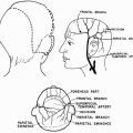
Since the marginal vessels are relatively large for such a small flap, its viability is extremely good, despite 180° rotation of the flap. It is important to realize that the marginal eyelid vessels lie about 3 mm from the lid margin and immediately beneath the layer of the orbicularis muscle.
OPERATIVE TECHNIQUE
Defects of Up to Half the Upper Eyelid
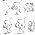
Because of the degree of stretch in eyelid tissues (that permits defects of up to a quarter of the lid to be closed directly), it is necessary to take a flap from the lower lid of not more than one-quarter of its width (approximately 6 to 7 mm). Hence the lower lid can be closed directly (Fig. 23.1) after the division of the vascular pedicle 2 weeks later.
The marginal vessels should not be damaged. Moreover, the pedicle width should not be less than 5 mm, and the various wounds must not be closed under tension. For reconstructions of this size, the hinge of the flap should lie directly below the center of the upper lid defect. It can be placed on either side, whichever is more suitable, always remembering that the lower lid punctum should never be sacrificed.
Defects Between Half and Three-Quarters of the Upper Eyelid
If the defect in the upper lid is a few millimeters greater than half the upper lid width, the lower lid flap will be a few millimeters larger than a quarter of the width of the lower lid (Fig. 23.2). This small additional segment of lower eyelid must be reconstructed by moving a small lateral cheek rotation flap lined with conjunctiva or with a composite nasal septal graft of cartilage and mucosa (see Chapter 13).
Related posts:
 Cheek Rotation Skin (Mustardé) Flap to The Lower Eyelid
 Wraparound Cartilage Flap for Correction of Cleft-Lip Nasal Deformity
 Oral Mucosal Flaps for Septal Reconstruction
 Postauricular and Retroauricular Scalping Flap (The Paras Flap)
 Scapular and Parascapular Flaps
 Platysma Musculocutaneous Flap to The Lower Lip
Stay updated, free articles. Join our Telegram channel

Full access? Get Clinical Tree


