Skin Island Orbicularis Oculi Musculocutaneous Flap for Lower Eyelid Reconstruction
R. V. ARGAMASO
A substantial loss of lower eyelid tissue may result from trauma or surgery. Reconstruction by skin-flap transfer has been of particular interest to reconstructive surgeons.
INDICATIONS
In recent years, the introduction of musculocutaneous flaps has increased the survival rate of skin transfers. In many instances, the quality of results also has improved.
It is noteworthy that long before musculocutaneous flaps became popular, Tripier performed a transposition of an orbicularis oculi musculocutaneous flap from the upper to the lower eyelid in 1889 (1). The same concept was elaborated on for repair of lower lid defects after tumor ablation (2). The Tripier flap was further modified by converting the skin component into an island on a bipedicled orbicularis oculi muscle that was then transferred to the lower lid for correction of severe ectropion (3).
Described below is a skin island orbicularis oculi musculocutaneous flap obtained from the suprabrow area and used for reconstruction of the lower eyelid. The upper eyelid was not a suitable donor, since it was also partially damaged.
ANATOMY
The orbicularis oculi is a flat, circular muscle arising from the nasal part of the frontal bone, frontal process of the maxilla, and anterior surface of the medial palpebral ligament. The pretarsal and preseptal sections occupy the eyelids, while the orbital portion surrounds the orbits, spreading to the temple and down to the cheeks. The muscle layer is relatively thinner within the eyelids than that which surrounds the orbits.
The latter forms a complete ellipse without interruption. Its upper fibers blend with those of the frontalis and corrugator muscles.
The latter forms a complete ellipse without interruption. Its upper fibers blend with those of the frontalis and corrugator muscles.
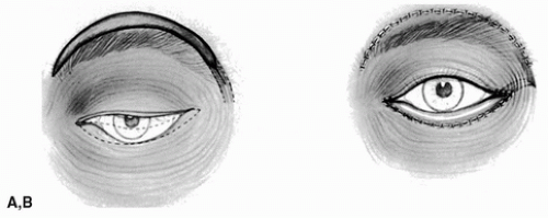 FIGURE 18.1 A: Diagram of the skin island outlined above the eyebrow. The flap should be as wide as the defect of the lower lid. Broken lines delineate the muscle pedicle. B: The skin island is sutured in place. The muscle pedicle, actually wider than the width of the skin island, lies in a subcutaneous tunnel.
Related posts: Cheek Rotation Skin (Mustardé) Flap to The Lower Eyelid Cheek Rotation Skin (Mustardé) Flap to The Lower Eyelid
 Wraparound Cartilage Flap for Correction of Cleft-Lip Nasal Deformity Wraparound Cartilage Flap for Correction of Cleft-Lip Nasal Deformity
 Oral Mucosal Flaps for Septal Reconstruction Oral Mucosal Flaps for Septal Reconstruction
 Postauricular and Retroauricular Scalping Flap (The Paras Flap) Postauricular and Retroauricular Scalping Flap (The Paras Flap)
 Scapular and Parascapular Flaps Scapular and Parascapular Flaps
 Platysma Musculocutaneous Flap to The Lower Lip Platysma Musculocutaneous Flap to The Lower Lip
Stay updated, free articles. Join our Telegram channel
Full access? Get Clinical Tree
 Get Clinical Tree app for offline access

|