Mohs micrographic surgery has the highest cure rate for skin cancer. Accurate and precise preparation of horizontal frozen sections in the laboratory is essential for the success of Mohs micrographic surgery. Key considerations in developing the Mohs surgery laboratory are careful planning and design, selection of proper equipment and supplies, training of laboratory personnel, adherence to regulatory standards of Clinical Laboratory Improvement Amendments (CLIA), and execution of an effective daily routine. The method of tissue processing used in the laboratory must yield optimal results for processing skin in an efficient manner.
Mohs micrographic surgery has the highest cure rate for skin cancer, while preserving the surrounding normal tissue. The success rate of the procedure is dependent on microscopic examination of 100% of the surgical margin at the time of surgery. Accurate and precise preparation of horizontally cut quality frozen sections in the Mohs laboratory is essential. A thoughtful approach is needed when designing a successful, efficient, and safe Mohs laboratory. The essential categories of development of the laboratory are planning and design, selection of proper equipment and supplies, training of laboratory personnel, adherence to regulatory standards of Clinical Laboratory Improvement Amendments (CLIA), and execution of an efficient and accurate daily routine.
Design of space
The Mohs laboratory should be located close to the surgical suites to allow ease of transporting tissue from the patient’s room to the laboratory. Grossing and inking of the Mohs specimen may take place in the surgical suite or the laboratory. Grossing and inking of the tissue in the patient’s room is often beneficial for ensuring anatomic accuracy of mapping and inking of the specimen. A wheeled cart that can be readily moved from room to room may be used for the grossing and inking process in each room.
The laboratory should be organized to follow the flow of the specimen processing. Division of the laboratory into a workbench area and a microscope viewing area is helpful. Bench height laboratory stools and 91.5-cm (36-inch) countertops are used in the workbench area during the processing of tissue. Desk height stools and 76-cm (30-inch) countertops are placed in the microscope viewing area for the surgeon. Having a separate designated area within the laboratory for the surgeon’s microscopic examination of the frozen sections is recommended. The surgeon’s close proximity to the Mohs technician allows ease of communication regarding special considerations with specimen processing and need for recuts. A 2-headed microscope is helpful for teaching purposes. These microscopes are available with a side-by-side adapter and a face-to-face adapter. Counter space and laboratory design should be constructed according to the type of adapter that is used. With the advent of electronic medical records, it is helpful to have a computer easily accessible for the technician in the laboratory.
The specimens arrive in the laboratory and are placed on an open countertop near the cryostat. The cryostat must be located in an easily accessible area. A separate 20-A dedicated circuit is required. Two cryostats are often placed in the laboratory to have a backup cryostat should the main cryostat malfunction. Avoid placing the cryostat directly under an air vent, which could lead to problems with maintaining temperature and frost accumulation. A microscope near the cryostat and staining area is helpful for preview of the slides by the Mohs technician before staining. This process provides immediate feedback to the technician about the completeness of the epidermal edge in the sections. Additional sections can be taken at this point, as needed, rather than waiting for evaluation by the surgeon after staining.
Adequate chemical-resistant countertop space is needed for staining. Manual staining requires a linear arrangement of Coplin jars or staining dishes in an area with adequate ventilation and a fume hood, if hazardous chemicals are used. An automatic stainer can be fitted with an appropriate-sized fume hood. A fume hood with a back draft is available to fit under overhead cabinets, if required. The presence of a fan-powered exhaust vent that exhausts directly to the outdoors also helps to decrease any chemical odors generated in the laboratory.
A flammable liquid storage cabinet should be located in the laboratory for storage of flammable chemicals. The metal cabinet should be labeled clearly and placed away from exit doorways.
Other considerations for the laboratory are designated areas for biohazard disposal, an eyewash station, slide storage, supply storage, textbooks and other references, and a sink for hand washing near the exit.
Equipment
Basic equipment needs for the Mohs laboratory are the cryostat, staining equipment, and a microscope. The cryostat is the largest single purchase for the laboratory. The most commonly used brands of cryostats are made by Leica Microsystems (Bannockburn, IL, USA): TBS Minotome Plus (Triangle Biomedical Systems, Raleigh, NC, USA) and Microm. The Microm cryostat has recently been replaced by the Avantik QS11 (Avantik Biogroup, Springfield, NJ, USA). Key components of a cryostat are the microtome, cryochamber, blade holder, handwheel, and freeze plate. Features for consideration are a motorized versus manual object head, Peltier cooling, ultraviolet disinfection, antiroll plate, and heat extractor. The cryostat should be kept between −20 and −25°C. Two or 3 cryostats may be desirable based on the volume of cases and need for a backup cryostat. If you do not have a backup cryostat, CLIA require you to have documentation of your plan of action if your cryostat quits functioning.
An automatic stainer can reduce reagent waste, increase efficiency in the laboratory, and enable more consistent staining. A survey by Robinson in 2001 reported that use of the automatic stainer, the Linistainer system, for routine slide preparation decreased staining variability by approximately 20% and reduced processing time by about 30%. Other benefits of the automatic stainer were cited as increased technician availability for mapping tissue, increased caseload, ability to perform larger cases with more sections, and performing other laboratory techniques. The most commonly used stainer is the Linistat (Thermo Scientific, Kalamazoo, MI, USA), which is 63.5 cm (25 inches) long. The average time for staining is less than 5 minutes. The stainer requires a supply line and drain tubing that may simply run into a sink. It may be connected directly under the sink, so that the sink can still be used for hand washing or for an attached eyewash. A fume hood is recommended over the automatic stainer. Supplies for manual staining are also recommended.
The microscope for the surgeon’s examination of the prepared sections is the other major purchase for the Mohs laboratory. Recommended features of the microscope for reading of the slides include a 2 to 2.5× low-power screening objective, 4×, 10× and 40× objectives, ceramic stage, 5-position turret, long-life bulb, flat field optics, and mechanical stage tension adjustment control. A tilting binocular tube can be helpful to prevent back and neck issues. As mentioned earlier, a dual-view side-by-side adapter or dual-view face-to-face adapter may be desired for teaching purposes.
Other equipment needs are a microscope for use by the Mohs technicians for reviewing slides for quality and completeness before presenting them to the surgeon, cryogen, any special embedding equipment, fire extinguisher, safety storage cabinet for flammable materials, and an eyewash station. A slide storage system is necessary to store biopsy and positive Mohs section slides indefinitely and negative Mohs section slides for 10 years. All slides should be saved for at least 10 years.
Supplies
An evolving list of supplies is useful to ensure adequate inventory and minimize excess. Box 1 outlines the commonly needed supplies in the Mohs laboratory. The reagents vary based on the desired staining protocol. However, generally the following reagents are necessary: 95% alcohol, 100% alcohol, xylene or xylene substitute, hematoxylin, eosin, bluing reagent, optimal cutting temperature (OCT) compound or freezing matrix, cover slipping glue, positive charged slides, cover glass, and gauze. If you use disposable blades, it is suggested you keep an extra pack on hand. When ordering supplies, order several reagents at a time because there is a hazardous shipping charge on all chemicals shipped; this charge is the same whatever the amount of the shipment.
25-mm specimen chucks
30-mm specimen chucks
Scalpel
Forceps
Microtome lubricating oil
Microtome tools
Antiroll brushes
Diposable blades
Specimen storage tray
Charge slides
Coverslips
Petri dishes
Marking dyes
Cryospray or liquid nitrogen
Tissue freezing medium
OCT compound
Mounting medium
Reagents for staining
Slidefolders
Slide storage system
Specimen processing
Several different methods of tissue processing in Mohs surgery have been developed through the years. The method of tissue processing must give optimal results for processing skin, including the epidermis, dermis, and adipose, in the least amount of time. Some of the described techniques include heat extractor, glass microscope slide, Bard Parker scalpel handle, Miami Special (Delasco, Council Bluffs, IA, USA), Cryomold (Sakura Finetek, Torrance, CA, USA), forceps, American Optical tissue presser, and the cryoembedder (Cryoembedder, Salt Lake City, UT, USA).
When processing tissue during a Mohs procedure the cryostat needs to be turned to −21 to −25°C. The micrometer setting is best set about 6 μm for skin sections. At this setting you can achieve a webbing effect in fatty specimens.
The tissue is brought to the laboratory in a secondary container along with a completed Mohs map. The map gives the location and a diagram of the Mohs layer. The map also contains the gross description of the specimen submitted to the laboratory. The Mohs map is marked with the time and technician’s initials when the tissue arrives in the laboratory. The tissue may be processed as 1 section or divided, as needed. Several different methods have been described to aid in obtaining a complete margin and epidermal edge in frozen sections, such as beveled excision, ex vivo or in vivo relaxing incisions, mechanical tissue flattening, and division of the tissue. The tissue may be carefully scored around the edges to place all the epidermis and deep margin completely down on the same plane. After the scoring is complete, the technician or surgeon dyes the tissue to ensure proper orientation when the surgeon is reviewing each section under the microscope. The dye and orientation are marked on the map for future review, and they must correspond correctly with the tissue so the surgeon can identify the location(s) of the remaining positive margin.
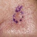
The surgeon marks the tissue with a notch at 12 o’clock to maintain orientation; the tissue is marked with red dye to the right of the notch at 12 o’clock, blue dye to the left of the 12 o’clock margin, and green dye at 6 o’clock; if needed, yellow dye and black dye can be added for layers containing several quadrants. All quadrants should have at least 3 focal points under the microscope, as seen in Fig. 1 .
Each quadrant has its own differential markings. This method of inking is used so that if the tissue is flipped, it cannot be confused with another quadrant.
After the tissue has been marked with the dyes, the tissue is ready to be embedded. There are several techniques to embed the tissue or prepare the tissue to be sectioned in the cryostat. Three of the more commonly used techniques are discussed.
The first technique is the slide technique. The tissue is placed deep side down on the slide, making sure all skin edges are flat against the slide ( Fig. 2 ). A little OCT compound is placed around the tissue and this is snap frozen in liquid nitrogen (LN 2 ) for approximately 10 to 15 seconds. It may be placed in a small cup of LN 2 or frozen on the quick-freeze bar of the cryostat. There should be a little graying left on top of the OCT compound ( Fig. 3 ). The specimen on the slide is then slightly thawed in the palm of the technician’s hand and the button is slid off the slide ( Fig. 4 ). A little OCT compound is placed on a chuck in the cryostat, and the button from the slide is inverted and placed on the chuck with OCT compound in the cryostat. The flat side is now in the up position, with the tissue ready for sectioning ( Fig. 5 ). The heat sink that is in the cryostat is placed on top of the newly formed button and allowed to freeze. The chuck is ready to section in seconds ( Fig. 6 ).
Related posts:
 Mohs Micrographic Surgery Technique
 Mohs Surgery for Squamous Cell Carcinoma
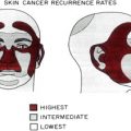
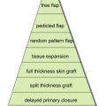 Flaps and Grafts Reconstruction
 Management of Unusual Cutaneous Malignancies: Atypical Fibroxanthoma, Malignant Fibrous Histiocytoma, Sebaceous Carcinoma, Extramammary Paget Disease
 Nonsurgical Treatment of Nonmelanoma Skin Cancer
Special Considerations for Mohs Micrographic Surgery on the Eyelids, Lips, Genitalia, and Nail Unit
Nonsurgical Treatment of Nonmelanoma Skin Cancer
Special Considerations for Mohs Micrographic Surgery on the Eyelids, Lips, Genitalia, and Nail Unit
Stay updated, free articles. Join our Telegram channel

Full access? Get Clinical Tree


