Secondary Syphilis
John T. Crissey
(ICD-9 091.3)
Symptoms and Signs
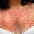
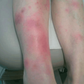
The lesions of secondary syphilis (SS) are discrete asymptomatic, erythematous to copper-colored, macular, and maculopapular lesions, which present on the trunk and genitalia. A prodrome of sore throat and flu-like symptoms may precede the lesions by a few days. The face is usually heavily involved, especially in the seborrheic areas and along the hair line. Brownish or coppery macules and slightly scaly papules often appear on the palms and soles (Fig. 25-1). More than any other feature, these lesions should alert the examiner to the possibility of syphilis.
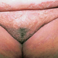
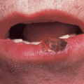
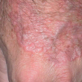
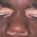
In other cases, SS lesions have a pronounced tendency to assume annular configurations, particularly in black patients. The face, anogenital areas, palms and soles, axillae, and periumbilical areas may be involved. Because of their size and configuration, these lesions are often called “nickel and dime syphilids” (Fig. 25-2). Mucous membrane lesions include a transient diffuse redness of the throat and the so-called mucous patches. The latter are slightly elevated round or oval papules, 5 to 12 mm in diameter, faintly inflammatory and covered with a pearly or grayish membrane. Lesions at the labial commissures may take the form of “split” or fissured papules, easily confused with ordinary perlèche. Mucocutaneous lesions in the genitalia and anal areas may appear as condylomata lata, which vary in morphology from slightly pedunculated, flat papules to smooth and button-like lesions or large cauliflower-like vegetations.
Related posts:
Stay updated, free articles. Join our Telegram channel

Full access? Get Clinical Tree


