63
Scapholunate Instability
Steven F. Viegas and Manuel F. DaSilva
History and Clinical Presentation
A 45-year-old right hand dominant railroad worker fell at work onto his right hand. He developed pain in the wrist, a painful snapping sensation, and weakness of pinch and grip strength in that hand. He was treated with an extended period of splinting and nonsteroidal antiinflammatory medications without success. Subsequent radiographs demonstrated a scapholunate dissociation with dorsal intercalated segment instability (DISI) deformity. Wrist arthrogram demonstrated a disruption in the scapholunate interosseous ligament. The patient was referred for further treatment 9 months after the injury.
Physical Examination
The patient had localized tenderness at the dorsal aspect of the scapholunate joint. Scaphoid stress test resulted in pain and an audible and palpable snapping. Range of motion of the wrist was full and comparable to the contralateral side. Pinch and grip strengths were ∼45% of the contralateral side.
Physical examination should include both extremities; unless otherwise indicated, the contralateral side is an excellent measure of the patient’s normal baseline regarding range of motion and level of physiologic laxity. When attempting to assess whether or not scapholunate dissociation exists, one should pay particular attention to any tenderness at the level of the scapholunate joint, particularly at the dorsal aspect of the scapholunate joint, on average located approximately 1 cm distal to Lister’s tubercle. The Watson test or scaphoid stress test is also useful to assess whether or not there exists any abnormal mobility of the scaphoid. Occasionally, a painful, audible snap can be elicited during this test.
Diagnostic Studies
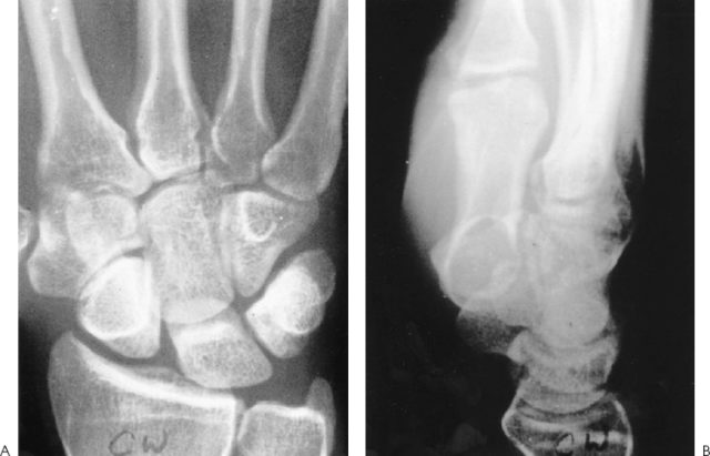
Radiographs demonstrated a foreshortened scaphoid with a positive signet ring sign with an increased scapholunate joint space on an anteroposterior (AP) view (Fig. 63–1A). There was a DISI alignment on the lateral radiograph (Fig. 63–1B). Previous wrist arthrogram demonstrated communication between the midcarpal and the proximal wrist joints through the scapholunate joint.
Appropriate radiographs should be obtained, including standard posteroanterior (PA), lateral, radial deviation, ulnar deviation, oblique, and AP supinated clenched fist views of the wrist and hand. Radiographs of the contralateral wrist can be obtained and used for comparison. A significant scapholunate dissociation will result in a carpal instability dissociative (CID) type of lesion that typically will result in a DISI pattern of carpal malalignment. This carpal instability pattern has been well described regarding its particular radiologic features, which include the following:
PEARLS
- Identify visually and/or by palpation the location of the DIC ligament before making the initial capsular incision.
- Anatomic reduction of the scapholunate joint is critical and needs to be confirmed visually from both the mid- and radiocarpal joints as well as radiographically.
- It is important to understand the lateral “V” construct, which consists of the dorsal radiocarpal ligament, distal intercarpal ligament, and dorsal components of the scapholunate and lunotriquetral ligaments, that allows indirect dorsal stability of the scaphoid throughout its range of motion.
PITFALLS
- Wrists with lunotriquetral coalitions can have a widened scapholunate joint space without having a scapholunate dissociation.
- A patient can have a positive arthrogram showing dye leakage through the scapholunate joint and/or arthroscopic identification of a tear of the membranous portion of the scapholunate ligament without having scapholunate instability.
- Use of an inadequate number and/or size of K wires may result in pin breakage or bending or suboptimal immobilization of the carpus.

- Scapholunate gap: The intercarpal distance between the scaphoid and lunate on the AP radiograph is increased, compared with the other intercarpal spacing. A scapholunate gap greater than 3 mm is considered diagnostic of a scapholunate dissociation. This scaphoid gap has been called the “Terry Thomas” sign after the famous English film comedian’s dental diastema. This increase in the scapholunate intercarpal distance is most noticeable, both in biomechanical and clinical studies, in the AP supinated clenched fist radiographic view.
- A shortened scaphoid: The scaphoid assumes a flexed posture as a result of its dissociation from the surrounding carpus and assumes a shorter appearance on the PA and AP radiographic views.
- The cortical ring sign: The flexed posture of the scaphoid results in an end-on view of the scaphoid tubercle/distal scaphoid, which results in this more prominently visualized circular cortex of the scaphoid.
- DISI pattern of the carpus: The scaphoid assumes a flexed and dorsally subluxed posture, the lunate assumes an extended and volar subluxed posture, and the capitate lies in a flexed posture.
- Taleisnik’s “V” sign: The intersection of the volar edge of the scaphoid outline when the scaphoid is flexed intersects with the volar margin of the radius at a more acute angle than the normal wrist in which there is a more gentle or wide C-shaped pattern of intersection.
Stay updated, free articles. Join our Telegram channel

Full access? Get Clinical Tree


