69
Scapholunate Advanced Collapse
Andrew H. Borom and David B. Siegel
History and Clinical Presentation
A 54-year-old right hand dominant man who works as a museum director presented with a 3-year history of gradually increasing pain, swelling, and loss of mobility in his right wrist. He also noted the recent onset of nocturnal paresthesias in the sensory distribution of the median nerve. There was no history of trauma, although he had played professional basketball for 12 years. A wrist splint, nonsteroidal antiinflammatory medication, and therapeutic modalities did not relieve his symptoms.
PEARLS
- The status of the articular cartilage on the proximal surface of the capitate is critical. Radiographic and intraoperative findings must be reconciled, and a decision must be made at the time of surgery as to the procedure chosen.
- In both operative procedures (proximal row carpectomy and 4-bone arthrodesis) a partial denervation of the wrist is indicated by excision of the terminal branch of the posterior interosseous nerve in the floor of the fourth dorsal compartment.
- Leaving a thin volar shell of bone avoids violation of the palmar extrinsic radiocarpal ligaments and does not appear to compromise the result.
PITFALLS
- Failure to reduce and stabilize the capitolunate joint in the four-bone arthrodesis will compromise outcome and reduce postoperative extension secondary to radiocapitate impingement.
- The pins used to stabilize the intercarpal fusion should not cross the radiocarpal joint.
- Silicone implants to replace the scaphoid are contraindicated, and biologic interposition material adds nothing to success.
Physical Examination
The right wrist had an effusion and was tender over the radial styloid-scaphoid joint. Range of motion was restricted to less than 50% of the contralateral side with complete loss of radial deviation. Extension and radial deviation were most painful. A scaphoid shift test provoked pain but no scaphoid subluxation. Provocative signs for carpal tunnel syndrome were positive; however, there was no thenar atrophy.
Diagnostic Studies
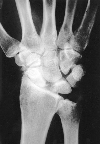
Radiographic imaging included posteroanterior, lateral, and scaphoid oblique views. There was narrowing of the joint space between the radial styloid process and the scaphoid, widening of the scapholunate joint, malrotation of the scaphoid, and narrowing of the capitolunate joint space (Fig. 69–1). There was beaking of the radial styloid process. Electrical diagnostic studies confirmed the presence of carpal tunnel syndrome.
Differential Diagnosis
Posttraumatic osteoarthritis
Scapholunate advanced collapse
Scaphoid nonunion or malunion with collapse
Distal radius fracture with radiocarpal collapse
Inflammatory arthritis
Rheumatoid arthritis
Gout
Diagnosis
Scapholunate Advanced Collapse Pattern of Osteoarthritis
Chronic scapholunate dissociation leads to a predictable pattern of degenerative changes in the radiocarpal and midcarpal joints. The incongruent joint surfaces between the radius and malrotated scaphoid, as well as those between the head of the capitate and dorsiflexed lunate, lead to osteoarthritis. The radiolunate joint is preserved, as it maintains a congruent relationship despite the rotatory malalignment. Joint deterioration follows a predictable sequence with well-described radiographic findings.
Figure 69–1 Preoperative radiograph demonstrates collapse of the radioscaphoid joint, marginal osteophyte formation at the distal tip of the radial styloid process, and mild narrowing of the capitolunate joint.
Stay updated, free articles. Join our Telegram channel

Full access? Get Clinical Tree


