CHAPTER 29 Rotation Mastopexy

Summary
The rotation mastopexy is a technique based on an anatomical approach.1 The lower half of the breast gland, carrying the nipple–areola complex is raised as a large flap based on a superomedial vascular supply. This is rotated in a craniolateral direction through 90° placing lateral glandular tissue into a pocket underneath the upper pole, so augmenting the upper pole with autogenous tissue, which is well vascularized. Excess skin is excised without tension and closed in a vertical fashion.
Key Points
Patient Selection
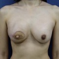
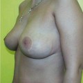
Using Regnault’s2 classification this technique is suitable for patients with Grade 1, Grade 2, and Grade 3 ptosis. After breastfeeding and with aging, the breast tissue drops to a varying degree with associated skin and ligamentous stretch. The breast also flattens and widens with some bulk of the breast tissue sitting laterally. The aim of the mastopexy is to restore tissue to the upper pole and create a breast mound with a round shape and projection (Figs 29.1–29.3). This should be done discarding a minimal amount of tissue in order to retain as much volume as possible.
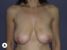
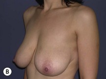
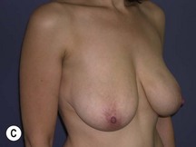
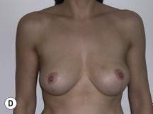
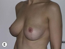
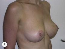
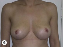
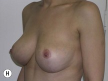
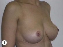
Fig. 29.1 Rotation mastopexy.
With permission from Corduff N, Taylor GI. Rotation mastopexy: an anatomical approach. Aesth Plast Surg 2009;33:377, Springer.
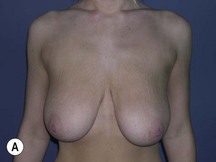
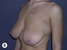
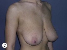
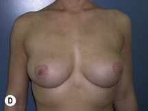

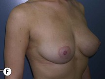
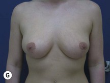
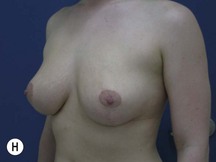
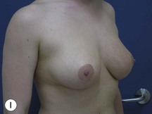
Fig. 29.2 Rotation mastopexy.
With permission from Corduff N, Taylor GI. Rotation mastopexy: an anatomical approach. Aesth Plast Surg 2009;33:377, Springer.
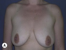
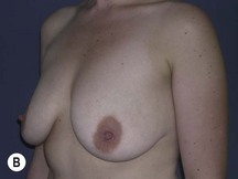
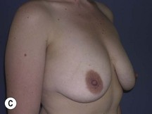
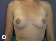
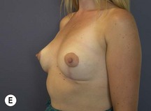
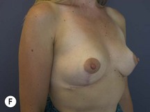
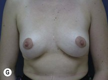
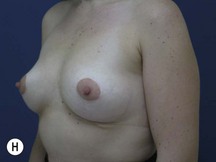
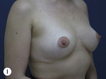
Fig. 29.3 Rotation mastopexy.
With permission from Corduff N, Taylor GI. Rotation mastopexy: an anatomical approach. Aesth Plast Surg 2009;33:377, Springer.
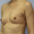
The technique is also suited to those patients who have breast ptosis in association with breast implants in situ (Fig. 29.4). In a patient who has had breast implants in situ for many years it is not unusual to find the tissue in the central and upper poles to be thinned out, with the bulk of the breast tissue to be lying below the implant. Many of these patients have put on some weight and there is sufficient volume to recreate an adequate breast mound from the available tissue without having to use new implants.
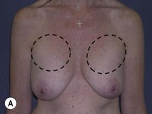

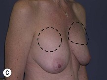
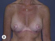
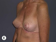
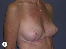
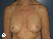
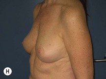
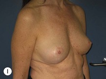
Fig. 29.4 Removal of implants with rotation mastopexy.
With permission from Corduff N, Taylor GI. Rotation mastopexy: an anatomical approach. Aesth Plast Surg 2009;33:377, Springer.
Indications
In restoration of the breast shape to a more youthful appearance, volume needs to be restored to the upper pole. The skin envelope is reduced and the nipple restored to an aesthetically correct position on the new breast mound. Commonly, adaptation of breast reduction techniques3 are used in a mastopexy, resecting mainly skin and a smaller amount of breast tissue. Following surgery the tightened skin envelope relaxes over time and the upper pole fullness is often lost leaving a less than optimal result. Ideally a technique should be used that reshapes the breast without relying on the skin envelope to hold it. Techniques have been described using small glandular flaps transposed to increase projection,4–9 but these do not adequately fill out the upper pole. Alternatively combining the reduction approach with an implant to fill out the upper pole10–14 is well described but associated with a significant complication rate.13,14
In those patients for explantation of breast prostheses, mastopexy at the time of implant removal is often a good option in that it will avoid a secondary deformity. Otherwise there is a risk that the pocket will contract down in an irregular fashion and lead to a subsequent deformity that can be hard to fix, especially in a patient whose tissues are not as elastic as they once were.15
It has been suggested that leaving the capsule behind can lead to increased seroma formation.16 This has not been the experience of the author. The approach to explantation is to leave the capsule behind if it is not heavily calcified and there is no free silicone present around the implant. Otherwise as much of the capsule that can be safely removed is excised. (Often some of the posterior capsule in the subpectoral pocket is left where it is closely adherent to the ribs.)