(1)
Department of Dermatology, Freiburg University Medical Center, Freiburg, Germany
Abstract
In some genodermatoses, one or more mosaic patches of healthy skin may result from revertant mutations occurring during postnatal life. – Molecular evidence of revertant mosaicism has been provided in the autosomal dominant traits, epidermolysis bullosa of the Dowling-Meara type and ichthyosis with confetti (ichthyosis variegata). Additional clinical examples are cases of allelic didymosis as reported in Darier disease and epidermolytic ichthyosis of Brocq. – Moreover, revertant mosaicism was proven at the molecular level in several autosomal recessive traits such as non-Herlitz junctional epidermolysis bullosa, dystrophic epidermolasis bullosa, and Kindler syndrome. Sometimes, even two or three different back mutations were found in the same individual. Such findings have fostered the hope that new strategies of gene therapy, avoiding the problem of viral vectors, may be developed.
In both autosomal dominant and recessive skin disorders, a thorough clinical examination may reveal some patches of healthy skin reflecting revertant mutations. Moreover, in some Mendelian disorders such as ichthyosis in confetti (ichthyosis variegata) or Kindler syndrome, multiple events of mitotic recombination or slipped mispairing may give rise to numerous patches of healthy skin [5, 9, 11]. In these traits, manifold reversion mechanisms appear to be rather the rule than an exceptional event.
11.1 Revertant Mosaicism in Autosomal Dominant Skin Disorders
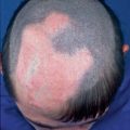
Patches of clinically healthy skin with molecular evidence of revertant mosaicism were documented in epidermolysis bullosa of the Dowling-Meara type [16] and in ichthyosis variegata (ichthyosis with confetti) (Fig. 11.1) [2–5]. Moreover, clinical evidence for revertant mosaicism without molecular examination was described in epidermolytic ichthyosis of Brocq [6] and Darier disease [7, 15] (see Sects. 3.1.1.6, 8.1.3, and 8.1.4).
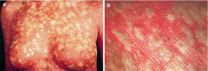
Fig. 11.1
(a) Ichthyosis variegata (ichthyosis with confetti) showing thousands of islands of revertant mosaicism [3] (Reprinted with permission from the British Association of Dermatologists); (b) Close-up showing hypertrichosis of the areas affected by the forward mutation [2] (Reprinted with permission from Karger Publishers, Basel, Switzerland)
11.2 Revertant Mosaicism in Autosomal Recessive Skin Disorders
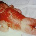
Patches of skin that did not blister have been noted in non-Herlitz junctional epidermolysis bullosa (Fig. 11.2) [13, 14], recessive dystrophic epidermolysis bullosa [1, 14], and Kindler syndrome [9, 11] (Fig. 11.3) (see Sect. 3.1.1.6). In all of these cases, molecular analysis revealed diverse events of revertant mosaicism.
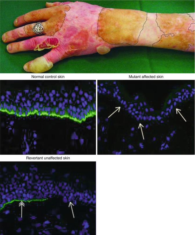
Fig. 11.2


Revertant mosaicism in a case of recessive junctional epidermolysis bullosa. Biopsies showed absence of collagen 17 staining (green) in the mutant affected skin, whereas patchy re-expression of the protein was noted in the revertant unaffected areas [14]. Note that the patient prefers to wear her ring on a finger with revertant unaffected skin (Reprinted with permission from Discovery Medicine, Publisher of Johns Hopkins CME Series)
Related posts:

Stay updated, free articles. Join our Telegram channel
Full access? Get Clinical Tree
