Reverse Temporalis Flap for Reconstruction After Orbital Exenteration
N. MENON
R. SILVERMAN
EDITORIAL COMMENT
The temporalis muscle is an excellent local flap to fill the exenterated orbit. The authors’ suggestion of making an adequate orbitotomy to pass the muscle has the advantages of shortening the arc of rotation and avoiding an osteotomy of the lateral orbital rim. The secondary depression in the temple can be filled as the authors indicate with either allograft or dermal-fat grafts.
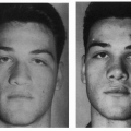
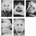
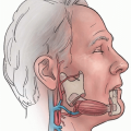
Orbital exenteration usually involves the resection of the entire contents of the orbits, with or without resection of the eyelids. Techniques to replace the orbital contents with vascularized tissue have involved multiple stages or have included the use of microvascular techniques. This chapter introduces the reader to a single-stage pedicled transposition of the temporalis muscle into the orbital cavity through a window created in the lateral orbital wall.
INDICATIONS
Techniques to replace the orbital contents with vascularized tissue have included pedicled flaps (retroauricular island flap, cheek flap, pectoralis flap) (1, 2, 3) and free flaps (radial forearm, dorsalis pedis flap) (4, 5, 6). A single-stage pedicled transposition
of the entire temporalis muscle as a pedicled flap into the orbital cavity, through a window created in the lateral orbital wall, can be accomplished in any patient who has undergone orbital exenteration and has an intact ipsilateral temporalis muscle. This single-stage technique provides the muscle bulk needed to obliterate the orbital cavity, and provides a vascularized soft-tissue cover around the exposed bone and subjacent sinus cavities.
of the entire temporalis muscle as a pedicled flap into the orbital cavity, through a window created in the lateral orbital wall, can be accomplished in any patient who has undergone orbital exenteration and has an intact ipsilateral temporalis muscle. This single-stage technique provides the muscle bulk needed to obliterate the orbital cavity, and provides a vascularized soft-tissue cover around the exposed bone and subjacent sinus cavities.
ANATOMY
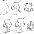
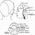
The temporalis muscle is a fan-shaped muscle located on the lateral aspect of the skull. It originates along the inferior temporal line on the cranium. The deep temporal fascia covers both the superficial and the deep (cranial) aspects of the muscle. The deep temporal fascia joins the periosteum of the cranium along the superior temporal line (temporal crest). The muscle lies in the temporal fossa and inserts on the mandible after passing along the deep surface of the zygomatic arch (7). The muscle inserts on the coronoid process and along the anterior ramus of the mandible. The muscle is innervated by the mandibular branch of the trigeminal nerve (cranial nerve V). Contraction of the muscle causes retraction and elevation of the mandible (7).
The muscle is supplied by two dominant vascular pedicles and a minor pedicle, making it a type III muscle (Mathes and Nahai classification) (7). The dominant pedicles are the anterior and posterior deep temporal arteries, both of which are branches of the internal maxillary artery. The deep middle temporal artery (branch of the superficial temporal artery) forms the minor vascular pedicle (2). The venous drainage of the muscle is through the venae comitantes that accompany the arterial supply.
Both dominant vascular pedicles are located on the deep surface of the muscle at the level of the zygomatic arch within a centimeter of each other. On the other hand, the minor pedicle is located on the superficial surface of the muscle in the preauricular region just cephalad to the zygomatic arch (7).
Related posts:
 Cheek Rotation Skin (Mustardé) Flap to The Lower Eyelid
 Wraparound Cartilage Flap for Correction of Cleft-Lip Nasal Deformity
 Oral Mucosal Flaps for Septal Reconstruction
 Postauricular and Retroauricular Scalping Flap (The Paras Flap)
 Platysma Musculocutaneous Flap to The Lower Lip
 Microvascular Free Transfer of Serratus Anterior and Rib Composite Flap
Stay updated, free articles. Join our Telegram channel

Full access? Get Clinical Tree


