Retroauricular Dermal Pedicled Skin Flap to The Ear
A. J. RENARD
Techniques described to correct defects of the external ear usually fall into two categories. The first includes circumference-reducing procedures with the necessary removal of normal tissue and a resulting small ear (1, 2, 3, 4). The second includes various techniques designed to maintain the size of the ear by interposition of a flap or graft or both (5, 6, 7, 8, 9). The use of a retroauricular flap for reconstruction of the auricle through an opening in the cartilage is well known. The flap described in Chapter 88 uses this technique; however, it is based on a rather nonmobile subcutaneous pedicle (10).
ANATOMY
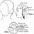
This is usually a random-pattern skin flap (11, 12). It is not uncommon to find a direct cutaneous artery at its base (Fig. 90.1). When this is the case, this retroauricular flap is an axial-pattern flap, receiving its blood supply in a retrograde fashion by the anastomosis between the superficial temporal artery and the retroauricular artery (Fig. 90.2).
FLAP DESIGN AND DIMENSIONS
The retroauricular dermal pedicled skin flap is designed as an ellipse (instead of a triangle) with its long axis oriented slightly obliquely, downward, and forward (Fig. 90.3). This orientation is important because as the donor defect is closed, the base of the flap is in a position that avoids distortion and undue tension on its dermal pedicle (Fig. 90.4). The base of the flap is deepithelialized over a length of about 1.5 to 2 cm. The greatest width usually measures approximately 2 to 3 cm (Fig. 90.3A).
Related posts:
 Cheek Rotation Skin (Mustardé) Flap to The Lower Eyelid
 Nasalis Musculocutaneous Sliding Flaps for Nasal Tip Reconstruction
 Inferior Turbinate Flap for Closure of Septal Perforations
 Postauricular and Retroauricular Scalping Flap (The Paras Flap)
 Scapular and Parascapular Flaps
 Platysma Musculocutaneous Flap to The Lower Lip
Stay updated, free articles. Join our Telegram channel

Full access? Get Clinical Tree


