Fig. 11.1
Typical assessment pathway. CT-PET, computed tomography-positron emission tomography; MRI, magnetic resonance imaging
Those considered appropriate for pelvic exenteration surgery should then be consulted with the decisions made at the multidisciplinary meeting. This allows for informed consent to be obtained in an unhurried and scheduled manner. The issues are complex when a patient and their family have to consider a large ablative operation compared with the alternative management options. These patients require considerable consultation and psychosocial support and counseling. Consenting patients are then sent for assessment before surgery. This process involves the following:
Anesthetic review and work-up (chest radiograph and electrocardiogram with or without a pulmonary function test, blood gas measurement, echocardiogram, and respiratory/cardiology assessment)
Stoma therapy assessment
Nutrition and dietary assessment
Psychosocial assessment, support, and counseling, during which patients are offered patient-to-patient contact with those who have already undergone pelvic exenteration surgery
Table 11.1 demonstrates the operative and admission details for patients undergoing pelvic exenteration surgery.
Median operating time | 9 h (range, 3–16 h) |
Median blood transfusion rate | 6.6 units (range, 0–17 units) |
Mean number of specialties involved | 3 (range, 1–5) |
Median number of days in intensive care unit | 3 days (range, 0–11 days) |
Median number of days in hospital | 25 days (range, 5–126 days) |
Patients should be discharged to the intensive care unit (ICU) immediately for initial postoperative care. On average, patients spend approximately 2–3 days in the ICU after pelvic exenteration surgery. In the ICU, daily care should involve:
The intensive care team
Primary care surgical review by colorectal surgeons
Surgical specialty review (urologist/plastic surgeon/orthopedic surgeon/vascular consultant, fellow, registrar, resident)
Stoma therapy
Nursing and allied health staff (dietician, physiotherapist, total parental nutrition nurse specialist)
Of note, patients who undergo sacrectomy or a vertical rectus abdominus myocutaneous (VRAM) flap to reconstruct the perineal or sacral defect should be nursed in a 30° lateral tilt position to prevent pressure on the surgical site. Upon stabilization, the patient can then be transferred to a ward with nursing and allied health staff that has experience in looking after these patients. On average, pelvic exenteration patients spend 22 days on the ward before being discharged to rehabilitation facilities. The following activities should occur on a daily basis:
Review by colorectal team
Review by surgical specialty team (urologist/plastic surgeon/orthopedic surgeon/v consultants, fellows, registrars, residents)
Nursing care (these patients need intensive management and often require two nurses at a time to care for each patient)
Stoma therapy
Allied health staff (dietician, physiotherapist, total parenteral nutrition nurse specialist, social worker, chaplaincy)
For the first 3 months after discharge from the hospital, patients should attend the outpatient clinic on a monthly basis or more frequently if necessary. All medical and allied health staff involved in the patient’s care should review the patient at this time. After this period, when patients are well enough, they can be transferred back to their referring specialist for continued outpatient care every 6 months. At a minimum, all patients should be followed up with a serum carcinoembryonic antigen level. CT scanning can be difficult to interpret after reoperative surgery because abnormal anatomy and radiotherapy make it difficult to differentiate scar tissue from tumor recurrence. CT/PET or MRI/PET scanning would be a more useful radiological follow-up tool because combined PET imaging can help elucidate between scar tissue, abnormal anatomy, and tumour recurrence.
Classification of Pelvic Exenteration Surgery
Locally recurrent rectal cancer is a difficult and complex management challenge. The aim of surgery is perform an R0 resection, removing all malignant or cancer tissue with a perimeter of normal tissue from the pelvis. Pelvic exenteration surgery may involve removal of the bladder, prostate, uterus and fallopian tubes, vagina, rectum, pelvic vessels and nerves, or bony components of the pelvic bone (e.g., sacrum) en bloc with the malignant disease. This radical surgery can leave the patient with a large tissue defect that requires reconstruction and repair with large myocutaneous tissue flaps.
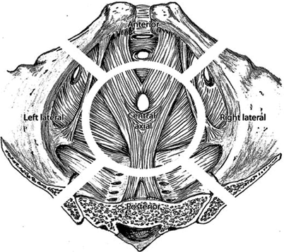
Patients who present with locally recurrent rectal cancer are a heterogeneous group in terms of the involved pelvic structures, and as such the definition of the extent of resection is debatable and individualized. As a result, there is no standard defined surgical procedure that is performed, but instead the type of operation is dependent on the site and size of the tumor and the number of organs involved. To help understand the operative approaches, the pelvis can be divided into four main compartments, which are geographically indicative of the nature of the pelvic recurrence (Fig. 11.2) [28, 29].


Fig. 11.2
Illustration of the anterior, axial, posterior, and lateral compartments of the pelvis
1.
The anterior compartment consists of the bladder, prostate, seminal vesicles, vas deferens, urethra, urogenital diaphragm, dorsal vein complex, obturator internus and externus muscles, anterior pelvic floor muscles (pubococcygeus and puborectalis part of the levator ani), pelvic bone (pubic symphysis, superior and inferior pubic rami), and obturator nerves and vessels.
2.
The axial compartment consists of the vagina, uterus, ovaries, fallopian tubes, broad ligament, round ligament of uterus, rectum, and pelvic floor muscles (iliococcygeus part of the levator ani).
3.
The posterior compartment consists of the rectum, pelvic floor (coccygeus muscle), internal iliac vessels branches and tributaries, piriformis muscle, sacral nerves S1-S4, pelvic bones (sacrum and coccyx), anterior sacrococcygeal ligament, and medial sacrotuberous and sacrospinous ligaments.
4.
The lateral compartment consists of the pelvic side wall structures, ureters, internal iliac vessels, external iliac vessels, piriformis and obturator internus muscles around the ischial spine, coccygeus muscle, lateral sacrotuberous and sacrospinous ligaments attached to the ischium, the ischium (including the ischial tuberosity and ischial spine), the lumbosacral trunk, and the sciatic nerve distal to ischial spine.
In broad terms, the compartments are best understood by appreciating their central anatomic points because there is some degree of overlap of their peripheries in each group. The central axis of the anterior compartment is the urethra, for the axial compartment it is the tip of the coccyx, for the posterior compartment it is the third sacral vertebra, and for the lateral compartment it is the ischial spines. The key surgical exenteration resection plane revolves around the excision of the entire “involved” compartment up to and including the bony margins.
In view of the heterogeneity of the types of resection, these resections are best defined as either partial or complete pelvic exenterations (PEs). A complete PE is defined as removal of the primary or recurrent tumor (with or without its attached bone) with all remaining pelvic viscera, that is, all four anatomic components of the pelvis. Partial PE is defined as removal of the primary or recurrent tumor (with or without attached bone) with en bloc resection of up to three anatomical components of the pelvis.
PE always involves an abdominal approach, usually with a perineal completion phase that can be performed in the lithotomy or prone position. Excisions of the anterior, axial, and lateral compartments are best performed through an abdominal approach combined with the perineal lithotomy position. Posterior resection of the sacrum from the fourth sacral vertebra (S4) down and the sacrospinous ligaments allows radical excision of the posterior pelvic floor, which is approached from the abdominal side and often is better visualized in this way rather than from a prone position.
Because of the nature of the sacroiliac joint attachment, involvement of the third sacral vertebra (S3) and above requires a prone approach unless only the anterior cortex of the midline bones of the fifth lumbar vertebra (L5) and upper sacrum need to be resected (this can be performed abdominally). Lateral, higher sacral and full vertebral excision of S2 and S3 requires the posterior prone approach.
Depending on the number and type of pelvic organs involved in the malignant process, the procedure requires a multidisciplinary team of highly skilled consultant surgeons from the surgical specialties of colorectal, vascular, urologic, orthopedic, and plastic and reconstructive surgeries. Colorectal surgeons predominantly perform the surgery, with other surgical disciplines being involved at the appropriate time. Specialist anesthetists and experienced theatre nursing staff also are required, and the procedure can take from 8 to 20 h, with a mean of 9 h. When a lateral compartment or neurovascular excision is required bilaterally, the mean operating time is extended to 12 h.
Surgical Approaches to Recurrent Rectal Cancer
Anterior Recurrence
Anterior pelvic recurrence may involve any of the anterior compartment structures. Mobilization of the posterior (total mesorectal [TME] or presacral plane) and lateral planes (ischial spine to the obturator internus muscle) should be performed before assessing the degree of anterior involvement and fixation to bony structures.
Involvement of the uterus, vagina, or both requires bilateral salpingo-oophorectomy, radical hysterectomy, and posterior or radical vaginectomy en bloc with tumor and the rectum. Depending on the degree of vaginal resection or perineal resection, primary closure with vaginal reconstruction or myocutaneous flap reconstruction may be required. Myocutaneous flap reconstruction is generally favored because the perineal wounds are typically large and because patients often have previously received radiotherapy. In addition, the level of recurrence will determine whether an ultralow anterior resection with colonic J pouch formation or a radical abdominoperineal resection is necessary.
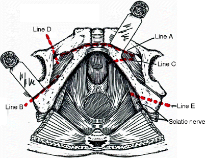
Involvement of the bladder dome or higher can be treated by partial cystectomy en bloc with the recurrent tumor and the rectum and with primary closure of the bladder. If the lower bladder (below the trigone), prostate, or both are involved, then radical cystectomy or cystoprostatectomy with ileal or colonic conduit formation is necessary. For anterior compartment tumors abutting or infiltrating the pubic bone, a more radical margin becomes necessary for the excision of the anterior regional structures. Wide exposure, but not incision, of the anterior levator muscles out to the inferior ramus of the pubic bone and back to the ischial tuberosity is performed from the perineum. The adductor and gracilis muscles are separated from their attachments to the lateral border of the inferior pubic rami and extended through the obturator fascia into the exposed pelvis. The inferior pubic ramus is transected free of the anterior pubis (line A) and posteriorly from the ischium (line B) with an oscillating or Gigli saw (Fig. 11.3).


Fig. 11.3
Lines of pubic bone transection
Excision of the pubic symphysis, if necessary, can be partial or complete en bloc with the tumor. The former is performed by bilateral exposure of the inferior pubic rami and their transection bilaterally (see line C in Fig. 11.3), separating it posteriorly from the ischial bones. Anteriorly, the inferior half of the pubis is horizontally transected below the superior pubic rami.
The superior half of the pubic symphysis combined with the superior pubic rami maintain stability of the pelvis. Removal of the entire pubic symphysis requires bilateral transection of the inferior (Fig. 11.3, line C) and superior pubic rami (Fig. 11.3, line D) as far laterally as possible. The pelvis is then structurally repaired using a polypropylene mesh to join the cut ends of all four pubic rami, which is then covered with a myocutaneous or rotation flap. The ischial tuberosity can be excised along line E (see Fig. 11.3), but care must be taken to protect the sciatic nerve as it passes posterior and lateral to the tuberosity. This should be conducted with the abdominal surgeon and perineal surgeon working in conjunction to identify and protect the sciatic nerve.
Central Recurrence
Central recurrence is otherwise known as an axial recurrence and usually includes anastomotic or “neomesorectal” recurrences. Patients frequently require an extended resection of surrounding tissues to ensure a clear circumferential resection margin. Thus, a radical abdominoperineal resection that takes a wide resection of the pelvic floor and perineum, including the S4 vertebra, usually is necessary. This can be performed with an osteotome at the level of the sacrospinous ligaments abdominally or, rarely, in the prone position. More proximal recurrences may be treated by an ultralow anterior resection and construction of a colonic J pouch; however, as a result of the heterogeneous nature of recurrent rectal cancer, these central recurrences often will involve resection of other organs including the uterus, fallopian tubes and ovaries, vagina, bladder, seminal vesicles, and prostate. When this scenario is encountered, it is treated as described earlier.
Lateral Recurrence
Lateral recurrence was previously considered a contraindication to pelvic exenteration surgery. The anatomic approach to lateral pelvic recurrence is in the plane lateral to the internal iliac vessels and necessitates en bloc excision of all or part of these structures. This plane allows access for en bloc dissection and excision of the lateral pelvic structures – the obturator internus and piriformis muscles, sacrotuberous and sacrospinous ligaments, and the sacral nerve roots – allowing access to the bony structures including the sacrum, ilium, or ischium, which also can be removed either from the anterior approach or in completion via a prone jack-knife position.
After a thorough abdominal exploration for metastatic disease and division of adhesions, dissection begins at the level of the aorta and inferior vena cava, with dissection and vessel looping of the ureters and common and external iliac vessels. At the level of the aortic bifurcation, the lymph node dissection is commenced and includes nodes overlying the aorto-iliac bifurcation and the common iliac and external iliac vessels until the origin of the internal iliac vessels is reached. The nodal tissue around the internal iliac territory is not entered and is taken en bloc with the vessels and specimen.


