Techniques to improve the chance of successful replantation of digits are well established. Indications and contraindications for replantation are generally agreed on, but they continue to evolve as excellent outcomes are achieved at centers with experience and expertise. Form and function can be restored with avulsion injuries and distal amputations, with good results and high patient satisfaction. Increased financial pressure to control the costs of health care and increased accountability for evidence-based outcomes may lead to the regionalization of replantation care and shared decision making in recommending replantation or revision amputation.
Key points
- •
Good outcomes (function, appearance, and patient satisfaction) in replantation are achievable with well-accepted techniques.
- •
The best outcomes are seen at high-volume, specialized centers with experience in replantation.
- •
To increase the likelihood that good outcomes will be achieved, regionalization of replantation care may need to be considered.
- •
To determine the most cost-effective treatment, more comparative studies of replantation and revision amputation need to be done.
- •
Although indications and contraindications for replantation are largely agreed on, the decision is shared between surgeon and patient. Anticipated function, costs, hospital stay, length of procedure, and time off from work should all be considered.
Introduction
Over the last 50 years, advances in microsurgical technique, bone fixation, nerve repair, and tendon repair have allowed salvage of amputated digits, hands, and limbs that would not have been possible in a previous era. Even in the earliest reports of these emerging and useful techniques, reconstructive microsurgeons stressed the importance of critically assessing function when examining outcomes. Simply considering survival of the replanted part is insufficient in determining the outcome and whether the combined efforts of surgeon, patient, and therapist are worth it. Range of motion, sensory recovery, and patient satisfaction all contribute to the overall outcome after replantation and should be evaluated, not just considering the successful reestablishment of blood flow and digit viability. Mechanism of injury (sharp, crush, or avulsion), level of injury (tip, relation to flexor digitorum superficialis, proximal interphalangeal joint involvement), and skill of the surgeon are all recognized as playing important roles in the overall outcome and function of replanted digits. Financial pressures are having an increasingly large impact on discussions regarding outcomes after replantation because of the costs of the procedures and time off from work for the patient. Evidence-based outcomes and cost accountability may lead to regionalization of hand trauma care, with patients being sent to centers with the highest volume and best outcomes.
This article reviews replantation of digits and addresses:
- •
Indications
- •
Accepted techniques and recent refinements (before, during, and after surgery)
- •
Outcomes after replantation
- •
New directions
Indications and contraindications
In deciding whether to reattach an amputated part, perhaps the most important step in the decision-making process is the discussion between the surgeon and patient about the time invested in treatment and rehabilitation, anticipated functional use, possible secondary procedures, and realistic expectations after a traumatic hand injury. The mechanism of injury, the level of injury, which digit is involved, patient occupation, time off from work, commitment to therapy, and accessibility to therapy are important in deciding whether to replant or perform a revision amputation.
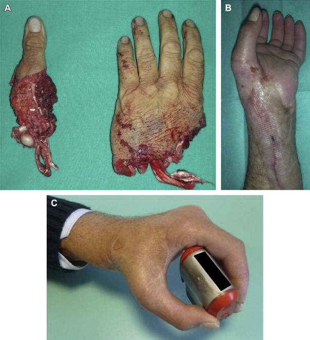
The most commonly accepted indications for replantation are thumb, digit in child, multiple digits ( Fig. 1 ), and distal to the flexor digitorum superficialis tendon insertion ( Table 1 ). There is disagreement with regard to the level of amputation, with some surgeons advocating revision amputation if the level is proximal to the flexor digitorum superficialis tendon insertion, whereas others have shown good functional outcomes with replantation at more proximal levels and involving only a single digit. Indications for replantation at very distal levels continue to evolve as good outcomes are reported in situations previously treated with revision amputation.
| Indications | Contraindications |
|---|---|
| Thumb | Extensive contamination |
| Digit in child | Comminuted fractures with significant bone loss |
| Multiple digits | Articular fracture with joint destruction |
| Level of amputation distal to flexor digitorum superficialis insertion | Multilevel injuries |
| Hand | — |
Factors that should be considered are patient age, patient comorbid conditions, ischemia time, level of injury, mechanism of injury, and severity of injury. Other traumatic injuries should be treated and take priority if life threatening. Accepted ischemia times for hand and digits are 6 hours and 12 hours for warm ischemia and 12 hours and 24 hours for cold ischemia, with reports of successful replantation after longer ischemia times (up to 33 hours for warm ischemia and up to 96 hours for cold ischemia).
Contraindications to replantation include extensive contamination, comminuted fractures with significant bone loss, and articular fractures with joint destruction (see Table 1 ). The mechanism of injury plays a significant role in whether an amputated part is suitable for replantation. A part that has been amputated sharply, in guillotine fashion, is more amendable to replantation than an avulsed part, which may contain injury to different structures at different levels. For example, the nerve and arterial injury may be at a different level than the tendon injury ( Fig. 2 ). Multilevel injuries make a good functional outcome much more difficult to achieve, and revision amputation is usually indicated.
Preoperative preparation
Transferring physicians from emergency rooms should be educated on the proper preservation of amputated parts as patients are transported to the treating facility. Wrapping the part in a saline-moistened gauze, putting in a plastic bag, and placing on ice is recommended. Placing directly on ice or in ice water should be avoided. Examination of the proximal amputation site and the amputated part is done to determine whether replantation is possible, based on the quality of the structures. Radiographs are also done to assess the extent of bony injury, with evaluation of the degree of comminution, intra-articular involvement, and bone loss. The decision to replant may require closer inspection of vessels under loupe magnification or using the microscope. Evidence of severe vessel injury, such as the ribbon sign or telescope sign, may make replantation impossible. If the vessel quality is acceptable, nerves, artery, and vein can be tagged with suture as the patient is prepared for surgery.
Intraoperative techniques
Sequence
Surgeon preference dictates the sequence of repair, with most advocating bone fixation first, followed by soft tissue repair of tendons, nerves, and vessels. Some surgeons prefer repair based on location of structures on the volar or dorsal sides, believing that repairs on one side should be completed before moving to the other side, to avoid excessive handling of structures and to prevent potential disruption of repairs. There is no consensus on the point in the sequence at which the veins are repaired. Some surgeons advocate repairing the vein before tourniquet release because of the bloodless field, whereas others advocate vein repair after reestablishment of arterial flow to allow easier identification of dorsal veins dilated with blood.
Bone
Bone shortening of 5 to 10 mm may be considered to provide better bone apposition for fixation if fractures are comminuted and to minimize tension on vessel and nerve repairs, especially when these vital structures are avulsed and excisional debridement is necessary. The options for bony fixation include Kirschner (K) wires ( Fig. 3 ), 90-90 intraosseous wires ( Fig. 4 ), screws, and plates. K wires are usually put in with the least difficulty and in the shortest amount of time, but internal fixation may make subsequent repair of structures in the sequence easier because internal fixation does not get in the way as much as K wires do. With intra-articular fractures, arthrodesis should be considered.
Tendon
After bone fixation or fusion, tendon repairs are done. Either standard Kessler sutures or pull-out sutures can be used. Both superficial and profundus flexor tendons are usually repaired.
Nerve
Nerve repairs can be done with the tourniquet inflated. Shortening of the nerve stumps may be needed to the level of healthier-looking fascicles. Dissection of the proximal and distal nerve ends should be done to mobilize the nerve as much as needed for a tension-free repair. Tissue glue or 8-0 or 9-0 nylon sutures can be used for repair. If a gap is present that prevents primary repair, nerve allograft, nerve autograft, neural tube, or vein graft conduit may be used.
Artery
Examination of the cut vessel ends under the microscope can reveal damage to the artery. Signs of severe vessel injury include the ribbon sign, the red line sign, the telescope sign, or the cobweb sign. Damaged vessel must be excised until the quality is suitable for anastomosis. Heparin solution is used to flush out the vessels. Vein grafts from the volar wrist can be harvested for arterial repair. A digital artery transfer from an adjacent digit may also be used rather than a vein graft. The tourniquet should be released to ensure good inflow from the proximal end. Traumatic amputation, vessel manipulation, and use of the tourniquet may lead to vasospasm, and arterial flow may not be seen after tourniquet release. Bathing the vessel in papavirine can induce vasodilation and demonstration of more robust inflow. Once good inflow has been confirmed and vessel ends are adequately trimmed and prepared, repair is done under the microscope with 10-0 or 11-0 nylon suture.
Vein
After arterial repair, observation of back bleeding of dorsal veins allows the selection of veins suitable for repair. If possible, repairing 2 veins for every 1 arterial repair has been recommended. Vein grafts may be required ( Fig. 5 ). In a traumatized digit, vein ends may be obscured by fat or debris. Thorough inspection of the subcutaneous plane under the microscope may be needed to identify suitable veins. Venous couplers or 9-0 or 10-0 nylon sutures are used for the repairs.
Related posts:


Stay updated, free articles. Join our Telegram channel

Full access? Get Clinical Tree


