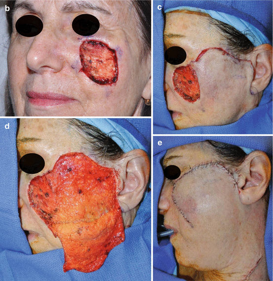
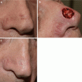
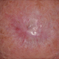
Fig. 10.1
(a–g) Left cheek lentigo maligna with final defect much larger than anticipated
Several studies have reported reconstruction techniques that were used following excision of LM/LMM. In the above noted study of 23 patients, 4 defects healed by secondary intention, 3 were closed primarily, 7 required skin grafts, 8 required local flaps, and 1 required a tissue expander with a subsequent flap [4]. In a study of 51 LM/LMM treated with staged excision, 36 % of patients were reconstructed with full thickness skin grafts, 22 % cervicofacial flaps, 14 % rhombic flaps, 12 % nasolabial flaps, 10 % rotation/advancement flaps, 4 % advancement flaps, and 4 % paramedian forehead flaps [5]. Temple and Arlette reported reconstruction techniques used in 166 LM/LMM patients treated with Mohs micrographic surgery. In this group, flaps were used in 59 patients, 48 had primary closures, 37 had skin grafts, 19 had a combination of grafts and flaps, and 3 underwent secondary intention healing [6].
With a mean age of 70 years [7], many patients with LM are elderly and may have multiple medical co-morbidities. Patient history including age, tobacco use, sun exposure, prior surgeries and a history of collagen vascular disease should be noted [8]. Exam should note skin laxity, location of langer’s lines, old scars and other nearby pigmented lesions that may need monitoring.
During initial patient evaluation it is important to remember that certain patients may not tolerate larger reconstructions that employ more distant flaps. A thorough discussion of patient expectations for cosmetic outcomes and complexity of the anticipated repair must occur.
A patient with limited life expectancy or co-morbidities that may preclude a longer procedure may not be physically capable or wish to undergo a more complex or multi-stage reconstruction. Although the cosmetic result may be sub-optimal, these patients should be considered for an office-based reconstruction with primary closure when possible, simple skin grafting, small local flaps, or healing by secondary intention [9].
Common Locations of LM
Lentigo maligna most commonly affects the cheek, followed by the nose [3, 10]. The two areas should be approached differently.
The cheek is a broad region extending to the lateral mandibular border. Asymmetries in the lateral region of the cheek are more forgiving than more central areas such as the nose or lips. Defects of the cheek are generally repaired by taking advantage of the laxity of local tissues without strict attention to subunits.
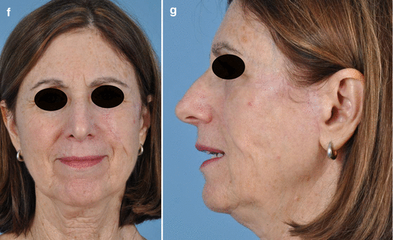
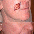
On the other hand, defects of the nose are typically closed with respect to exact subunit borders. Defects following margin-controlled excision techniques can be very broad and irregularly shaped. These defects may involve multiple cosmetic subunits on the face, requiring a combination of reconstructive techniques, as shown in Fig. 10.2a–g.
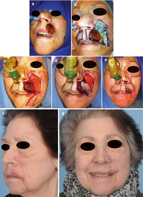
Fig. 10.2
(a–g) Complex upper lip defect requiring two separate advancement flaps
Wound Bed Preparation
After excision, it may be several days before negative margins are confirmed. In the meantime, the wound bed should be kept clean with saline gauze dressings, changed up to three times a day. Once negative surgical margins are histologically confirmed, reconstruction can commence.
The wound bed must be addressed first. It is important to clean the open wound and freshen the edges with a sharp blade, particularly if the reconstruction has been delayed.
Reconstruction
Reconstruction of any defect should be approached using standard principles and the reconstructive ladder.
Secondary Intention
While not often reported as the first choice for closure, in well-selected wounds, secondary intention can be a very useful and aesthetically appropriate method for healing. Particularly in the very elderly with multiple co-morbidities, healing by secondary intention may be the right approach, even in a site that is not cosmetically ideal. With proper selection of wounds, favorable aesthetic outcomes can be achieved following secondary intention healing. Specifically, concave wounds in locations such as the medial canthus, nasoalar sulcus, nasofacial junction, alar groove, temple, concha, and triangular fossa have shown excellent cosmetic results following secondary intention healing [11, 12]. Once the wound has healed and contracted, the residual scar that is much smaller than the initial defect can be serially excised. Figure 10.3a–d depicts the significant contraction demonstrated in a cheek wound that healed by secondary intention and has not yet undergone final reconstruction which will subsequently be a more minor procedure.
Fig. 10.3
(a–d) Medial cheek defect healed by secondary intention
Primary Closure
If healing by secondary intention is not reasonable, the next step is to assess how much potential local tissue advancement can be obtained by simply undermining the skin edges without violating any aesthetic subunits.
If a wound can be closed primarily on the cheek this should be done. Primary closure can match skin color, texture and thickness well. On the cheek it is crucial to ensure that a primary closure does not distort free margins such as the lower eyelid, causing ectropion [8].
Even if a primary closure can be achieved without free margin distortion, excessive tension on the wound should also be avoided. Undermining the soft tissues can create a fair amount of tissue recruitment especially in older patients with more skin laxity in order to achieve a tension free closure. Double-prong skin hooks should be used to retract the skin while undermining and sharp dissection is encouraged over bovie electro-cautery. Primary closure can be achieved by converting the defect into an ellipse, extending the length of the defect, but simultaneously avoiding dog ears. Another option is to utilize a purse-string closure for a circular defect; while the periphery of the wound may initially appear pleated, this effect resolves over time or could undergo a delayed secondary excision if necessary, working with a much smaller scar [13].
Skin Grafting
Although a skin graft does not always provide an optimal color and texture match, it is often the next best option for patients with significant medical co-morbidities who need stable coverage and cannot tolerate prolonged anesthesia. Skin grafts should not be used when there is exposed bone, nerves or blood vessels in the defect.
For cheek defects, a full thickness skin graft is preferred over a split-thickness graft. A full thickness graft provides a thicker substitute with a better texture match. They contract less over time, which is useful for near the eyelid, nose or oral commissure [8]. Donor sites for the cheek can include the axillary fold, supraclavicular region or pre-auricular region depending on the patient’s skin color and quality.
“Pie-crusting” of the graft (creating small holes in the graft for fluid egress) should be minimized for aesthetic reasons however a secure bolster is crucial for graft survival. Bolsters should remain in place for 5–7 days especially given that the cheek is such a mobile area. For defects involving the face, the surgeon should always use a bolster to secure the graft in place, as it is a difficult region that cannot be immobilized.
Local Flaps
A local flap can be appropriate for a moderately sized defect because it replaces “like tissue with like tissue” and therefore can camouflage well with time. All local flaps are designed so that at least one border of the defect becomes one side of the flap. These flaps take advantage of skin laxity adjacent to the defect to achieve closure. In older patients with deep relaxed skin tension lines the incision can be well hidden when properly placed. When approaching the cheek it is crucial to remember key pearls when creating flaps in the three different zones of the cheek, shown in Fig. 10.4.
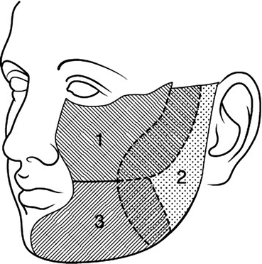
Fig. 10.4
The three zones of the cheek (Copyright ©2016, Memorial Sloan Kettering Cancer Center)
Zone I, the superior medial region, is amenable to hiding incisions in the naso-labial fold. Zone I often has less laxity than the other two zones. With the eyelid margin as the superior border, advancement of tissue should not be based inferiorly on the cheek to avoid lower eyelid pull and risk of ectropion.
Zone II, the pre auricular region, often has a fair amount of laxity in older patients, however care should be taken not to distort the beard or sideburns. Zone III is lateral to the mouth and extending down to the neck. Flaps in this area should not put any tension on the oral commissure, in this region, redundant tissue may be borrowed from the neck, below the mandibular border [14].
Related posts:
 Epidemiology and Natural History
 Incorporating Patient Preferences and Quality of Life
 Case B: Unsuspected Invasion and Upstaging in Lentigo Maligna Melanoma
 Staged Excision Techniques
 Follow Up and Recurrence
 Case A: Multiple Mapping Techniques to Guide Staged Excision for a Challenging Lentigo Maligna Melanoma
Stay updated, free articles. Join our Telegram channel

Full access? Get Clinical Tree


