18
Purpura and Disorders of Microvascular Occlusion
• As purpuric lesions fade, their color evolves from red-purple or blue to brown or yellow-green.
• Primary purpura has a broad differential diagnosis, and it is helpful to categorize purpuric lesions based on their size and morphology.
– Petechiae: ≤3 mm and macular (Table 18.1; Fig. 18.1A).
Table 18.1
Causes of petechiae and ecchymosis with minor trauma.
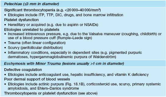
DIC, disseminated intravascular coagulation; ITP, idiopathic thrombocytopenia purpura; TTP, thrombotic thrombocytopenic purpura.

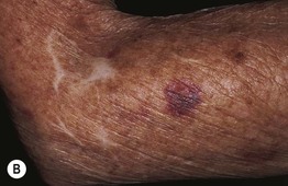
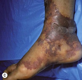
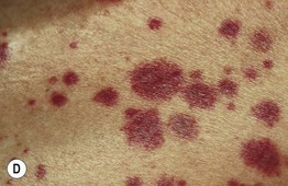
Fig. 18.1 Clinical examples of petechiae and purpura. A Round to oval petechiae, ≤3 mm in diameter. B Actinic (solar) purpura and pseudoscars, both in sites of actinic damage plus trauma. C Non-inflammatory (bland) retiform purpura as well as hemorrhagic bullae in a patient with disseminated intravascular coagulation (DIC). D Palpable purpura due to cutaneous small vessel vasculitis (inflammation plus hemorrhage). A, Courtesy, Warren Piette, MD; B, Courtesy, Jean L. Bolognia, MD; C, Courtesy, Judit Stenn, MD.
– Ecchymoses: usually >1 cm and macular with round/oval to slightly irregular borders, and typically have an element of trauma in their pathogenesis (see Table 18.1; Fig. 18.1B); a greater volume of hemorrhage leads to a hematoma, which is palpable.
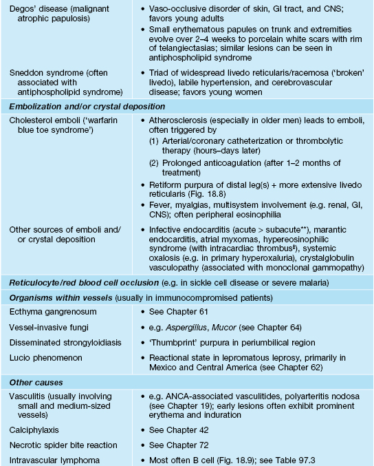
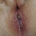
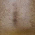
– Retiform purpura: reticulated, branching or stellate morphology, which reflects occlusion of the vessels that produce the livedo reticularis pattern (see Chapter 87; Tables 18.2 and 18.3; Figs. 18.1C and 18.2–18.9).
Table 18.2
Causes of retiform purpura.
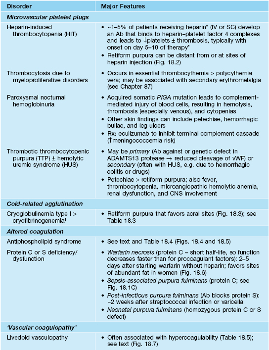
* Less common with low-molecular-weight heparin (≤1%) than unfractionated heparin; a transient decrease in the platelet count can also occur within the first 2 days of heparin therapy due to its direct effects on platelet activation.
† May be an incidental finding in hospitalized patients; cold agglutinins rarely lead to acrocyanosis or purpura.
** Skin lesions associated with subacute endocarditis are more likely to be inflammatory due to immune complex deposition, e.g. Osler’s nodes (tender red-purple papules), rather than Janeway lesions (purpuric macules; more common in acute endocarditis) on the hands and feet.
‡ Cutaneous microthrombi and superficial thrombophlebitis have also been described.
Ab, antibody; PIGA, phosphatidylinositol glycan anchor class A; vWF, von Willebrand factor.
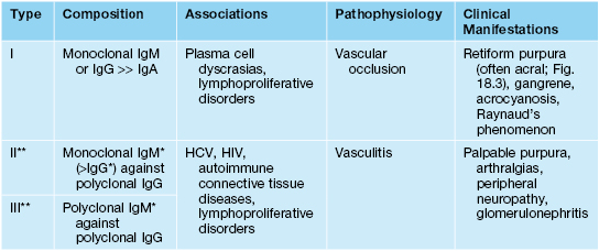
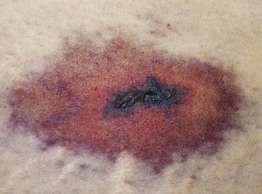
Fig. 18.2
Related posts:
Stay updated, free articles. Join our Telegram channel

Full access? Get Clinical Tree


