Fig. 45.1
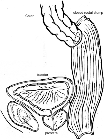
Introduction of the stapler through the anterior rectal wall in a “high” reconstruction. Note that the stapler is introduced at least 3–4 cm (depending on its size) distally into the rectal stump
This is especially applicable in cases where the Hartmann’s procedure was performed for benign disease. In cases where it was performed for cancer, particularly where there is any concern of local recurrence in the rectal stump, it is advisable to mobilize the rectum, to re-resect part of the rectal stump, and to make a final decision regarding reanastomosis depending on the findings of intraoperative histopathology. In such cases, an end-to-end anastomosis is advisable. In cases where an end-to-side anastomosis on the anterior rectal wall is considered (without the mobilization of the rectal stump), care should be taken to make the anastomosis at least 2 cm distal to the previous closure of the rectal stump because this area can be thickened, with reduced vascular supply (Fig. 45.2).


Fig. 45.2
A colorectal anastomosis is created on the anterior rectal wall in a “high” reconstruction. Note that the anastomosis is distant from the bladder and other pelvic organs
The surgeon should, when possible, be aware of the technique that was used for the rectal stump closure during the initial operation. If it was closed with a purse string (usually a double-layered suture), the stump tends to be thicker when compared with a stapled closure and anastomosis should be moved more distally. Likewise, in cases with excessive fibrosis in this area (due to previous suture leakage), the anastomosis should be created more distally to where the rectal wall is not thickened and has sufficient vascular supply. Anastomosis can be created manually or, preferably, with a circular stapler (single- or double-staple technique). It is not necessary (nor advisable) to make a purse-string suture on the anterior rectal wall when attempting a stapled anastomosis because simply passing the anvil of a circular stapler through a small hole in the rectal wall will be satisfactory in most cases. This hole should be created at least 3–4 cm (depending on the size of the circular stapler) distally to the rectal stump. We prefer curved, medium-sized (28–29 mm), circular staplers for this purpose because they can be more easily introduced through the rectum. The creation of an anastomosis on the anterior rectal wall simplifies the procedure, reduces operation time, and abolishes complications due to rectal stump mobilization.
The main avoidable complications include injury to the ureters (which generally are stented in malignant cases or in those where there has been prior radiation therapy), the gonadal vessels, and the hypogastric nerves, each of which may be adherent to the fibrous tissue of the rectal stump [30]. Preliminary computed tomography scanning in cases where ureteric stenting is contemplated provides anatomic information concerning the ureteric disposition (and number) as well as bilateral excretory function, which may influence this decision [31]. Ureteric stents are readily palpable during reconstructive surgery or they may be transilluminated. When there is apparent atrophy of the rectal stump (especially after longer delay of Hartmann’s reversal), there is a substantial risk of the rectal wall tearing during the introduction of a stapler. A full-thickness rectal wall tear represents a severe complication, but it can be easily identified during the procedure. More concealed, however, is tear of the rectal mucosa, which may also result in severe complications. These mucosal tears are easily overlooked until they become manifest with postoperative rectal bleeding, septic complications, or both. We believe that repeated balloon dilatation of the rectal stump or successive dilatation with rigid dilators 1 month before Hartmann’s reversal may be helpful in the prevention of this specific intraoperative complication. In these cases, a 28- to 29-mm circular stapler should be used. Measurement of the rectal diameter with rigid staple dilators before insertion of a stapler is strongly recommended to prevent rectal wall injuries. In cases where the introduction of a stapler is not safe after all these procedures, a hand-sewn anastomosis should be performed, if possible. The proximal part of the colon should be resected only a few centimeters above the colostomy and prepared for anastomosis. In case of residual uncomplicated diverticular disease proximal to the colostomy, that segment of the colon is resected if it is short in length and does not necessitate a major resection of colon. If the segment is long and would force a major colonic resection, it is advisable to leave it in situ, taking care not to incorporate some of the diverticular process into the new anastomosis. In most cases, there is no need for mobilization of splenic flexure to gain additional length of colon for a safe anastomosis without tension.
Mid Reconstruction
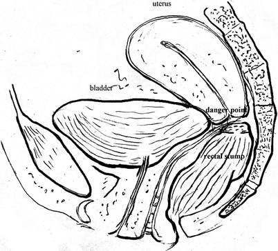
Mid reconstruction usually is indicated when the Hartmann’s operation was performed for rectal cancer. In these cases, the mesorectum was transected and the rectal stump closed deep in the pelvis. This kind of reconstruction is technically demanding and should be performed by an experienced colorectal surgeon. The surgical technique in this case is quite different from that of a high reconstruction. In most cases, especially in men, it is not possible to dissect a sufficient length of the anterior rectal wall to gain additional free space for the creation of a safe anastomosis, making dissection of the rectal stump necessary. In some women, dissection of the rectal wall from the vagina is possible, enabling the creation of an anastomosis on the anterior rectal wall. In these cases, care should be taken not to incorporate a part of the posterior vaginal wall into the anastomosis because this could result in the development of a rectovaginal fistula. However, dissection of the rectal stump, especially anteriorly, is challenging and prone to complications both in men and women; it should be avoided whenever possible and an end-to-end anastomosis is safer in these circumstances. Furthermore, the small bowel and the pelvic organs (bladder, uterus, ovaries, prostate, and seminal vesicles) tend to fill the empty space in the pelvis after rectal resection, making adhesiolysis particularly difficult (Fig. 45.3).


Fig. 45.3
The pelvic organs in women (bladder and uterus), which tend to fill the empty space in the pelvis after rectal resection, make adhesiolysis particularly difficult
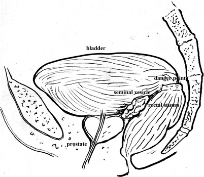
Difficulty specifically occurs in cases where the rectal stump was closed extraperitoneally without a complete closure of the pelvic peritoneum. We believe that covering the pelvic cavity with the peritoneum is an important step in the Hartmann’s procedure, making reversal easier and reducing the risk for small-bowel injury. However, by drawing the pelvic peritoneum forward during this procedure, both urethers may be brought medially if appropriate care was not taken to dissect them laterally before suturing. Dissection should always start from the mid-posterior side of the rectal stump and progress laterally because it is safer and less demanding. It should be emphasized that if dissection does not start from this position, both ureters may be transposed medially because of previous pelvic peritonealization, with considerable risk of injury. In cases with rectal stump dehiscence and a resolved/resolving pelvic abscess, adhesions between the rectal stump and surrounding organs may be thick and fibrotic, making adhesiolysis challenging. The risk of small bowel and bladder injury, especially in men, is high because the bladder tends to fall down into the pelvis and adhere to the rectal stump. Because of a narrow pelvis and lack of space, dissection in men is more difficult, with a high risk of bladder, seminal vesicular, prostatic, and pelvic nerve injury (Fig. 45.4).


Fig. 45.4
In men, the bladder tends to fall down into the pelvis and adhere to the rectal stump. Note that dissection in men is difficult because of a narrow pelvis and lack of space, with a comparatively higher risk for bladder, seminal vesicular, prostatic, and pelvic nerve injury
In women with a history of hysterectomy, there is a high risk of injury to the bladder and vagina as well as the risk of massive presacral bleeding. The latter complication is a specific feature of women who have undergone prior sacrocolpopexy, and presacral dissection is ill advised. Posterior dissection, if it is to be performed at all, should be conducted in the perimuscular plane (close to the rectal wall) whenever possible. Identification of the rectal stump and vagina by inserting metal dilators or fingers into the rectal stump and vagina can facilitate the difficult pelvic dissection. If the rectal stump can be positively identified after minimal dissection, the anvil of a circular stapler can be introduced through a small hole, which should be created toward the posterior rectal wall, especially in cases where the dissection of the anterior wall was difficult. Care should be taken not to incorporate parts of the bladder or vagina into the anastomosis. If an anterior dissection proves to be extremely difficult, with a high risk for injury to surrounding organs, the surgeon should focus on the posterior dissection and create an anastomosis on the posterior rectal wall. As stated earlier, although posterior rectal dissection is less demanding than anterior dissection, prior presacral dissection makes such posterior dissection particularly hazardous.
Low Reconstruction
Low reconstruction usually is indicated when the Hartmann’s operation was performed for mid-rectal cancer. In most cases, total mesorectal excision was performed and the rectal stump was closed deep in the pelvis at the level of the levator hiatus. This kind of reconstruction is demanding and should be performed only by an experienced colorectal surgeon. A short rectal stump, pelvic adhesions, and variable postoperative functional results are the main concerns when a Hartmann’s reversal is attempted in these cases. All previously described problems regarding pelvic adhesiolysis in mid reconstruction are present in low reconstruction but in a more severe form. After the removal of small-bowel adhesions from the pelvic cavity, the rectal stump dissection starts posteriorly. Surrounding organs should be meticulously dissected away from the rectal stump, with special care to prevent injuries to the bladder, prostate, and seminal vesicles in men and the uterus and vagina in women. Dissection of the rectal stump is facilitated by inserting a finger into the anal canal or the vagina. If the rectal stump can be dissected enough for safe anastomosis, end-to-end anastomosis with a circular stapler can be performed. In most cases, the rectal stump is thick, making anastomosis with a stapler relatively less safe. In such cases, a few radial incisions created at the apex of the rectal stump down to the muscularis propria may solve the problem; however, in most cases, safe dissection of such a short rectal stump is not possible and mucosectomy with a transanal hand-sewn coloanal anastomosis is recommended. With this technique, the extensive and dangerous dissection of the rectal stump is unnecessary. The rectal stump can be identified easily after complete transanal mucosectomy, which starts approximately 1 cm above the dentate line and ends at the apex of the rectal stump. Mucosectomy is aided by transabdominal and transanal maneuvers and is facilitated by submucous infiltration of diluted epinephrine to reduce bleeding and improve visibility and dissection in the correct plane. The transition zone should generally be preserved to improve functional results in accordance with the same technique originally described as part of ileal pouch anal anastomosis for inflammatory bowel disease or familial polyposis [32]. After complete mucosectomy, the rectal stump should be opened transanally through scar tissue under direct vision. The opening should be wide enough to accommodate the colon, which should be brought through the anal canal, followed by the creation of a coloanal hand-sewn anastomosis, as described by Parks [33]. We believe that a defunctioning loop ileostomy should be created at the end of this procedure. In most cases, the splenic flexure has to be fully mobilized with high ligation of the inferior mesenteric artery and vein to gain sufficient colonic length for the creation of a tension-free anastomosis. Splenic injury during mobilization of the splenic flexure is an infrequent but morbid complication. The reported rate of splenic injuries during colectomy is 0.42 %, rising to 3.1 % in cases with mobilization of the splenic flexure [34, 35].
Prior Continence Function
Prior continence function is an important issue, especially in cases for which low reconstruction is considered. It is well known that functional results after low anterior resection and coloanal anastomosis may be unsatisfactory, mainly because of fecal incontinence and evacuation disorders in the form of frequent and fragmented defecation (the so-called anterior resection syndrome) [36]. These problems may occur after Hartmann’s reversal, especially in cases with a low reconstruction, and patients should be fully informed about functional outcome before reversal. Routine evaluation of continence function is not necessary in most patients before reversal; however, patients with a history of minor incontinence or significant anorectal surgery should be evaluated. Low reconstruction is associated with a higher risk of fecal incontinence due to loss of the rectal reservoir, pelvic neuropathy, and potential sphincter damage caused by anal sphincter stretching during the creation of a transanal anastomosis. There is a group of patients, mostly women, with impaired continence function but without symptoms of fecal incontinence before the Hartmann operation. In most cases, they have an unrecognized obstetric sphincter injury or pudendal neuropathy.
After low reconstruction, the already damaged continence mechanism may not be sufficient in these new anatomic settings (loss of reservoir function of the rectum) and fecal incontinence may result. This group of patients is difficult to distinguish. Patients with questionable continence function should be selected and evaluated using questionnaires, anal manometry, and endoanal ultrasound. In cases where major incontinence can be expected, Hartmann’s reversal should be abandoned.
Evaluation of the Rectal Stump
The length of the remaining rectal stump is of paramount importance for defecatory function after Hartmann’s reversal because the rectum has a great capacity and serves as a reservoir for the stool before evacuation. The greater the amount of rectum resected, the greater the loss of rectal capacity and compliance, leading to urgency and frequent defecation. In addition, atrophy of the rectal stump and diversion colitis may occur in cases with a long period between the initial surgery and reversal, making functional results after reversal even worse. Fortunately, both rectal stump atrophy and diversion colitis can be reversed by reconstruction and should not preclude Hartmann’s reversal [37–39]. Routine endoscopic and contrast studies of the rectal stump frequently are performed before reconstruction, although there is a lack of evidence to support this practice [26]. In this respect, Cherukuri and colleagues [40] showed a low incidence of radiographic rectal stump abnormalities when patients were asymptomatic before Hartmann’s reversal, and most of the abnormal radiographic or endoscopic findings do not seem to influence definitive treatment [26, 41]. Our unit routinely utilizes preoperative radiography and endoscopy of the rectal stump, despite the specific value recorded by the literature, to define the length of the remaining rectum, the presence of unexpected leaks, strictures, inflammation, tumor recurrences, and atrophy of the rectal stump. This type of assessment is essential in cases where the Hartmann’s resection was performed elsewhere and may assist in operative planning and prediction of and counseling for anticipated functional outcome. In some cases, such information may provide the rationale for referral of some patients to a more experienced coloproctologist.
Functional Results and Quality of Life
The existing literature regarding Hartmann’s reconstruction mainly reports the short-term outcomes, including mortality and morbidity, of the procedure without discussing the quality of life or the functional results. Modern surgery recognizes the importance of standardized quality of life measures as crucial factors when assessing clinical outcomes after surgical interventions. Traditionally, it was believed that the quality of life was better after sphincter-saving procedures compared with permanent colostomies [42, 43], although many patient groups are not strictly comparable [44


Stay updated, free articles. Join our Telegram channel

Full access? Get Clinical Tree


