Pretibial myxedema


Pretibial myxedema


Cynthia O. Anyanwu, Preston W. Chadwick and Warren R. Heymann
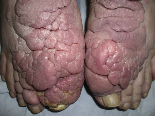

Evidence Levels:  A Double-blind study  B Clinical trial ≥ 20 subjects  C Clinical trial < 20 subjects  D Series ≥ 5 subjects  E Anecdotal case reports


image


Pretibial myxedema, more accurately termed ‘thyroid dermopathy,’ is characterized by non-pitting edema and skin-colored to violaceous nodules or plaques. These are most commonly distributed pretibially, but can sometimes be seen over the arms, shoulders, head, and neck.


Management strategy


Pretibial myxedema is an autoimmune phenomenon which tends to occur following treatment of patients with Graves disease. The condition can, however, develop in hypothyroid and euthyroid patients. It is helpful to look for other clinical signs of thyroid disease, including thyroid acropachy and the presence of a goiter. Pretibial myxedema typically follows the onset of ophthalmopathy, often years after the diagnosis of hyperthyroidism. Goals of treatment include cosmesis and the prevention of long-term side effects such as elephantiasis, decreased range of motion, or foot drop from neural entrapment. Resolution may occur without treatment.


Patients with significant thyroid dermopathy should be started on a trial of high-potency topical corticosteroids, alone or under occlusion, for at least 2 months. If symptoms persist, intralesional corticosteroids may be effective. A combination of the above in conjunction with compression bandages can be beneficial when monotherapy proves inadequate. Both oral and intravenous corticosteroids have also been shown to improve lesions in several patients. However, their use is limited by systemic side effects.


Pentoxifylline, an analog of methylxanthine theobromine, has been shown to reduce the extent of lesions and can also be used in conjunction with topical and/or intralesional corticosteroids. Although there are conflicting data, the use of intravenous immuneglobulin (IVIG) may improve lesions of pretibial myxedema. Subcutaneous or intralesional octreotide, a somatostatin analog, yields conflicting results. Plasmapheresis has been reported to be beneficial in improving severe cases and has been successful when used in combination with rituximab.


Temporary improvement with cytotoxic agents has been observed. Pretibial myxedema is not a life-threatening condition, and so the use of such agents should be limited to severe, debilitating cases. Surgical excision has been shown to be effective in a minority of cases. The high risk of recurrence makes surgical intervention an infrequently used modality; however, post-operative intralesional steroids can minimize this risk. Complete decongestive physiotherapy has shown some success in treating the elephantiasic form of pretibial myxedema.


Pretibial ultrasonography, with or without digital infrared thermal imaging, to measure skin thickness may be useful in assessing treatment response. Measuring serum hyaluronic acid levels to follow therapeutic response may also be of value.


Specific investigations


Observing pretibial myxedema in patients with Graves’ disease using digital infrared thermal imaging and high-resolution ultrasonography: for better records, early detection and further investigation.

Shih SR, Lin MS, Li HY, Yang HY, Hsiao YL, Chang MT, et al. Eur J Endocrinol 2011; 164: 605–11.


Digital infrared thermal imaging (DITI) detects surface temperature, and sonography reflects composition changes in soft tissue. Lower leg temperatures of normal volunteers decreased gradually from proximal to distal parts. In all patients with pretibial myxedema, DITI showed abnormally low focal temperatures over the lesions. In Graves disease patients with mild diffuse non-pitting edema and Graves disease patients with normal appearance of lower legs, DITI showed abnormally low focal temperature in 90.9% and 65.2% of the patients respectively. Areas of clinically visible pretibial myxedema and low focal temperature detected by DITI were sonographically characterized with increased skin thickness, hypoechoic substance deposition in the cutaneous tissue, and blurred boundary lines between dermis and subcutaneous tissue.


The use of digital infrared thermal imaging and high-resolution ultrasonography to analyze pretibial skin of patients with Graves disease allows for early detection of pretibial myxedema in patients with and without visible dermopathy.


Aug 7, 2016 | Posted by in Dermatology | Comments Off on Pretibial myxedema

Full access? Get Clinical Tree

Get Clinical Tree app for offline access