, Shimin Chang2, Jian Lin3 and Dajiang Song1
(1)
Department of Orthopedic Surgery, Changzheng Hospital Second Military Medical University, Shanghai, China
(2)
Department of Orthopedic Surgery, Yangpu Hospital Tongji University School of Medicine, Shanghai, China
(3)
Department of Microsurgery, Xinhu Hospital Shanghai Jiao Tong University, Shanghai, China
Zheng and Lin reported the anatomical basis and clinical application of posterolateral mid-forearm perforator flap [1, 2].
13.1 Vascular Anatomy
Three patterns of the perforator were observed in the posterolateral aspect of the mid-forearm [3].
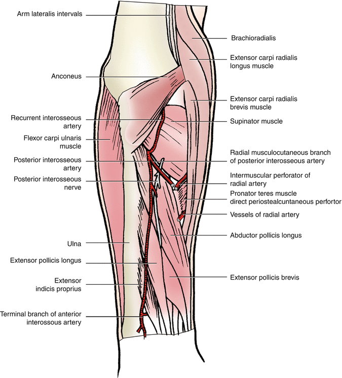
In pattern I, the perforator was the lateral terminal branch of the posterior interosseous artery that traveled initially between the contiguous border of supinator and abductor pollicis longus and gave off several muscular branches to the surrounding muscles along its course; the trunk of this perforator continued to emerge superficially at the septum between the extensor digitorum and extensor carpi radialis brevis and pierced the deep fascia at the posterolateral aspect of the mid-forearm. It pierced the deep fascia at the site 12.5 ± 0.3 cm inferior to the lateral humeral epicondyle along the line between the lateral humeral epicondyle and the radial head (Fig. 13.1).


Fig. 13.1
Vascular anatomy of the posterolateral mid-forearm perforator flap
In pattern II, the perforator originated from the proximal segment of the radial artery or the radial recurrent artery. It run initially at the septum between pronator teres and supinator in close contact with the radial periosteum and gave off several minute periosteal branches to nourish the middle segment of the radius. The trunk of this perforator traveled downward through the septum between extensor digitorum and extensor carpi radialis brevis and pierced the deep fascia at the posterolateral aspect of the mid-forearm. It pierced the deep fascia at the site 11.8 ± 0.2 cm inferior to the lateral humeral epicondyle along the line between the lateral humeral epicondyle and the radial head [4].
In pattern III, the perforator originated from the middle segment of the radial artery. It traveled obliquely downward along the inferior margin of pronator teres or traversed across the insertion of pronator teres, giving off several periosteal branches to form a vascular network in the mid-to-distal segment of the radius. The trunk of this perforator emerged superficially through the septum between extensor digitorum and extensor carpi radialis brevis, piecing the deep fascia at the posterolateral aspect of the mid-forearm. It pierced the deep fascia at the site 15.8 ± 0.4 cm inferior to the lateral humeral epicondyle along the line between the lateral humeral epicondyle and the radial head.
13.2 Illustrative Case
A 64-year-old male patient sustained a strangulation injury from a drilling machine at the forearm and wrist, leading to fractures of the radius and ulna, a soft tissue defect of 8.5 × 4.0 cm in size over the dorsal aspect of the mid-to-distal forearm.
Stay updated, free articles. Join our Telegram channel

Full access? Get Clinical Tree


