, Shimin Chang2, Jian Lin3 and Dajiang Song1
(1)
Department of Orthopedic Surgery, Changzheng Hospital Second Military Medical University, Shanghai, China
(2)
Department of Orthopedic Surgery, Yangpu Hospital Tongji University School of Medicine, Shanghai, China
(3)
Department of Microsurgery, Xinhu Hospital Shanghai Jiao Tong University, Shanghai, China
Hurwitz [1] described the first posterior thigh flap in 1980, when he used a musculocutaneous flap based on the inferior gluteal artery.
Ramirez et al. [2] reported on the posterolateral fascia lata flap, showing that it received branches from the first deep femoral perforating artery.
In 1984, Song et al. [3] documented a posterior thigh flap based on the third perforating branch of the profunda femoris artery.
In 1989, Maruyama and Iwahira [4] documented a popliteoposterior thigh fasciocutaneous island flap based on a direct branch of the popliteal artery for coverage of a knee wound.
This branch, however, was found to be inconsistent by Cormack and Lamberty [5].
In 1993, Paletta et al. [6] performed a posterior thigh flap based on the descending branch of the inferior gluteal artery to raise a posterior thigh island flap, an extended gluteal thigh flap, an ipsilateral pedicled thigh flap, and a local rotation flap.
In 1996, Lambert et al. [7] performed a posterior thigh fasciocutaneous flap based distally on a branch from the popliteal artery.
22.1 Vascular Anatomy
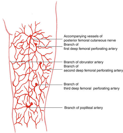
The largest part of the posterior thigh skin territory is supplied by the profunda femoris artery [5, 8], mostly by the first and second profunda femoris perforating arteries [9].
The first perforating branch of the profunda femoris artery arises superior to the adductor brevis muscle and pierces the adductor magnus muscle 2–4 cm inferior to the ischial tuberosity.
The third perforating branch of the profunda femoris artery arises inferior to the adductor brevis muscle and deep to the tendon of the adductor longus muscle. The vessel passes posteriorly through the adductor magnus muscle and the short head of the biceps femoris muscle and then sends branches to the vastus lateralis muscle. The third perforating branch ultimately gives off the branch that passes along the lateral intermuscular septum to supply the lateral thigh skin and the posterolateral thigh musculature. After this branch supplies the biceps femoris muscle, it also perforates the deep fascia to supply the skin of the median aspect of the posterior thigh, the pedicle of which ranges in length from 5 [3] to 10 cm [5].
Venae comitantes are of similar caliber. The fourth perforating branch of the profunda femoris artery is also its end artery. It is usually small and pierces the adductor magnus muscle just proximal to the level of the hiatus of the adductor magnus. The vessel supplies muscular branches and may supply branches that ramify between the semimembranosus and semitendinosus muscles. Alternatively, the fourth perforating branch may supply a branch toward the subcutaneous tissue in the popliteal fossa, between the semimembranosus and biceps femoris muscles (Fig. 22.1).


Fig. 22.1
Vascular anatomy of the posterior thigh perforator flap
22.2 Illustrative Case
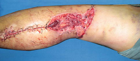
A 47-year-old man presented popliteal fossa defect measuring 14 cm × 6 cm after sustained traffic accident (Fig. 22.2).


Fig. 22.2
Preoperative view
Flap Design
Stay updated, free articles. Join our Telegram channel

Full access? Get Clinical Tree


