Posterior Labial Artery Flap for Vulvar and Vaginal Reconstruction
Chris A. Campbell
DEFINITION
The posterior labial artery flap commonly known as the Singapore flap or pudendal thigh flap is a thin fasciocutaneous flap oriented along the groin crease within the perineum.1
The flap’s pedicle, the posterior labial artery, is a branch of the internal pudendal artery system.
Unilateral or bilateral posterior labial artery flaps are commonly used for vulvar resurfacing and vaginal reconstruction.
ANATOMY
The posterior labial artery flap is inferiorly based with its axis along the groin crease including the thin skin lateral to the labia majora medially and the medial thigh skin laterally.
The flap’s pedicle is the posterior labial artery, which courses above the deep perineal fascia, arising from the perineal artery as it pierces the Colles (superficial perineal) fascia. The perineal artery branches off of the internal pudendal artery that emerges from the pudendal canal located 1 cm medial and inferior to the ischial tuberosity.
The flap’s skin paddle can be up to 15 cm long by 6 cm wide in the adult with the base of the flap even with the posterior edge of the introitus.2
The posterior labial branches of the pudendal nerve innervate the most posterior aspect of the skin paddle providing sensation to the reconstruction.
PATHOGENESIS
Vulvar or vaginal defects resulting from resection of squamous cell carcinomas, extensive condyloma, lichen sclerosis, fistulas, necrotizing soft tissue infections, and other cutaneous conditions have been reconstructed with unilateral or bilateral posterior labial artery flaps.
In the pediatric population, bilateral posterior labial artery flaps have been used for total vaginal reconstruction in cases of vaginal atresia.
PATIENT HISTORY AND PHYSICAL FINDINGS
When approaching vulvar or vaginal reconstruction, evaluate the abdomen, thighs, and inguinal region to maintain rectus and thigh-based flap alternatives to the posterior labial artery flap, in the event significant tissue bulk is required.
If the patient has had pelvic radiation to treat malignancy, the exam should focus on the quality of the groin and medial thigh skin to determine if it is within the radiated field. A severely radiated groin crease can increase the risk of partial flap loss and wound healing complications.
Measure the surface area of vulvar skin or vaginal mucosa that requires resurfacing to ensure that this amount of groin crease skin is redundant to allow for closure of the donor site.
IMAGING
Computed tomography of the pelvis will be part of the standard preoperative evaluation of patients requiring resection of vaginal abnormalities that will require subsequent reconstruction.
The posterior labial artery flap lacks bulk and as such is used to resurface the posterior or circumferential vagina or vulvar skin.3 Larger tumors that require pelvic exenteration will need alternative or additional flaps that provide soft tissue bulk in addition to skin resurfacing.
SURGICAL MANAGEMENT
Unilateral or paired posterior labial artery flaps are used to reconstruct partial or circumferential vaginal defects, respectively, and to resurface vulvar defects with thin pliable skin.
When total vaginal reconstruction is required, bilateral posterior labial artery flaps are sewn together and rotated into the pelvic defect producing a natural physiologic angle of inclination of the reconstructed vagina.4
Key considerations for use of the posterior labial artery flap include the ability to cover the vaginal or vulvar defect surface area with adjacent available groin skin within the design of the Singapore flap and whether the skin paddles are within fields of significant pelvic radiation.
Preoperative Planning
Pathology findings from prior biopsies and imaging studies should be reviewed to confirm the planned extirpation involving the vulva and/or vagina.
All resections that include the vagina will be preceded by an examination under anesthesia to determine if there has been disease progression and to confirm the planned resection and reconstruction.
A Foley catheter should be placed to protect urethral structures if total vaginectomy is required and to assist the patient with recovery.
Positioning
The patient is placed in the lithotomy position on the operating table for both the extirpative portion of the operation and the vulvar and/or vaginal reconstruction with one or both posterior labial artery flaps (FIG 1A).
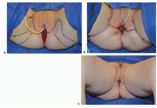
FIG 1 • Operative photos of bilateral posterior labial artery flaps for posterior vaginal reconstruction after rectal cancer resection. A. The patient is in lithotomy position with bilateral posterior labial artery flaps marked. Gluteal advancement flaps are also marked to obliterate the dead space from the abdominoperineal resection. B. Bilateral posterior labial artery flaps have been rotated into the pelvic defect to resurface the posterior and lateral surfaces of the vagina. C. Six months after surgery with donor-site incisions camouflaged within the groin creases and return of sexual function.Related posts:

Stay updated, free articles. Join our Telegram channel

Full access? Get Clinical Tree

 Get Clinical Tree app for offline access