Platelets are known primarily for their role in hemostasis, but there is increasing interest in the effect of platelets on wound healing. Platelet isolates such as platelet-rich plasma have been advocated to enhance and accelerate wound healing. This article describes the use of a novel preparation, platelet-rich fibrin matrix (PRFM), for facial plastic surgery applications such as volume augmentation, fat transfer supplementation, and as an adjunct to open surgical procedures.
Platelet-rich plasma (PRP) has been used clinically to simulate the native wound healing environment, but surgeons are cautioned not to generalize reported results; although many are approved by the US Food and Drug Administration, these preparations may vary greatly in erythrocyte contamination, leukocyte content, method of activation, and volume.
Platelet-rich fibrinogen matrix (PRFM) is a better product than PRP for use in facial plastic surgery because:
- •
The action of PRFM is more steady and sustained, yielding increased and sustained concentrations of growth factors during the crucial wound healing period after the initial acute inflammatory phase.
- •
The mechanical properties of PRFM, once fully polymerized, are significantly more stiff, representing a stiffness about half that of intact human skin.
- •
The robust scaffolding structure of PRFM possibly translates into resistance to physiologic stress, more accurate implantation, and presumably longer persistence and resistance to washout at the site of injection.
![]() V ideo of I njection T echniques with platelet – rich fibrin matrix accompanies this article at http://www.facialplastic.theclinics.com/ .
V ideo of I njection T echniques with platelet – rich fibrin matrix accompanies this article at http://www.facialplastic.theclinics.com/ .
Introduction
Platelets play a major role in hemostasis, but their functions in regulation of immune response, wound healing, osteogenesis, and angiogenesis have only recently become the subject of extensive investigation.
In vivo, activation of platelets is mediated by contact with the site of injury and attachment to the fibrin scaffolding formed at the site of injury, with subsequent biochemical cascades that lead to, among other effects, pseudopod formation, aggregation, and degranulation of platelets. Derived from megakaryocytes, platelets store bioactive molecules in their secretory organelles. α-Granules contain more than 300 proteins, many of which are yet to be characterized. These proteins are involved in many biologic roles including hemostasis and clotting, cell proliferation, extracellular matrix formation, angiogenesis, vascular modeling, chemotaxis, and inflammation. The complex interaction of these molecules with cells involved in wound repair, such as fibroblasts, macrophages, and endothelial cells, is central to understanding wound repair.
Some of the proteins released from α-granules of activated platelets are specifically involved in wound healing, including tumor growth factor β (TGF-β), platelet-derived growth factor (PDGF), insulinlike growth factor-1 (IGF-1), basic fibroblast growth factor (bFGF), vascular endothelial growth factor (VEGF), and connective tissue growth factor (CTGF). In addition, platelets release coagulation factors, serotonin, histamine, endostatin, and hydrolytic enzymes. As noted earlier, activation of platelets is mediated by contact with the site of injury and leads to the release of bioactive substances from platelets (mentioned earlier). The complex interaction of these molecules with cells involved in wound repair, such as fibroblasts, macrophages, and endothelial cells, is central to wound repair.
Growth factors are released in exact ratios and work in specific order, both independently and in concert, to lead to appropriate hemostasis, inflammation, and wound healing.
The use of exogenous growth factors
Therapeutically modifying the amount of these bioactive substances, and thus enhancing wound healing, is useful to the scientist and clinician. Pharmacologic agents such as human recombinant PDGF (becaplermin 0.01%, Regranex; Systagenix Wound Management, Inc., London, United Kingdom), in use for diabetic foot ulcers, and human recombinant keratinocyte growth factor used for oral mucositis in patients receiving chemotherapy (palifermin, Kepivance; Biovitrum AB, Stockholm, Sweden) have been formulated, studied, and shown to be effective.
In light of the abundance of bioactive chemicals in platelets and the complexity of their interactions and effects, studies of the application of single growth factors have not produced uniform or conclusive clinical results. In addition, exogenous growth factor application directly and outside a natural site of healing may have untoward effects: in 2008, the US Food and Drug Administration (FDA) issued a black box warning for the use of becaplermin, because patients exposed to 3 or more tubes of this drug had a 5-fold increase in cancer mortality. The safety of palifermin in patients with nonhematologic malignancies has not been established. However, if platelets can effectively be delivered to the site of injury, improved and accelerated healing may be expected.
Use of endogenous platelets
To better simulate the native wound healing environment, concentrated platelet preparations (PRP) have been used clinically. There are several FDA-approved systems to produce a PRP, but their products vary in erythrocyte contamination, leukocyte content, method of activation, and volume, and the reader is cautioned not to overly generalize reported results from PRP.
Platelet Preparations
Autologous blood is centrifuged followed by resuspension of platelets in a small amount of recovered plasma after erythrocytes and leukocytes are removed. This process yields a PRP with 3 to 5 times the normal concentration of platelets in peripheral blood. The PRP is then usually activated with calcium and bovine thrombin, which leads to platelet degranulation and massive release of all growth factors. Depending on the system used, some PRP preparations also contain leukocytes (predominantly lymphocytes); although there is some indication that leukocytes may enhance the antibacterial activity of PRP, they may be counterproductive in inducing tissue generation, because they are also known to release matrix metalloproteins and reactive oxygen species.
Surgeons in varied disciplines have used PRP to modulate wound healing, including attempts at accelerating the healing of bone grafts in orthopedic and sports medicine, as recently reviewed by Nguyen and colleagues and in the dental literature. PRP has also been used for improved healing of chronic lower extremity wounds and progressive hemifacial atrophy. Man and colleagues described the use of PRP in 20 patients undergoing cosmetic surgery including neck lift, face lift, breast augmentation, and breast reduction. Cervelli and colleagues reviewed the use of PRP in conjunction with fat grafting in several aesthetic and reconstructive procedures.
Clinical results reported with the use of PRP have been equivocal, possibly because most growth factors, such as TGF-b and PDGF, are released immediately from the PRP platelets, with significant reductions at days 3, 7, and 14. This finding may explain the transient effect of PRP on wound healing. In an animal study, Sclafani and colleagues noted an increase at day 7 in endothelial cells and fibroblasts after application of PRP to experimental wounds; however, this increase was lost by day 14. In a different experiment, Hom and colleagues used autologous platelet gel to treat wounds of the adult thigh, and although earlier wound epithelialization was noted with the use of the gel, ultimate cellularity was comparable with that of controls. This finding is supported by the work of other investigators, who found the effect of exogenous epidermal growth factor (EGF) application to be transient, and that only sustained application of EGF improved wound healing.
In their study comparing hemifaces treated with PRP before flap closure during deep-plane facelifts, Powell and colleagues did not note a significant difference in postoperative edema and ecchymosis compared with control hemifaces. Others failed to show any significant improvement with the clinical use of PRP in a randomized clinical study.
Platelet-Rich Fibrin Matrix
In addition to platelets and their products, the natural wound response requires the presence of a fibrin matrix, which enhances the delivery of growth factors. Fibrin mediates the adhesion of fibroblasts and other cells to the injured site. Furthermore, basic fibroblast growth factor (bFGF) has a high binding affinity specifically for fibrin and fibrinogen. Studies have shown enhanced survival and differentiation of transplanted preadipocytes when coinjected with fibrin as a carrier material compared with controls. Other clinical studies have reported good results when treating patients with autologous fat coinjected with PRFM.
Animal studies have also suggested improved wound healing when PRFM is used. Nitche and colleagues found that rabbit patellar tendon defects treated with surgery had more desirable wound healing when additionally treated with PRFM compared with surgical repair alone. This finding was quantified by decreased inflammation, more organized collagen deposition, and increased tensile strength at 3 weeks. This difference was not noticed at 6 weeks after surgery. In a different study, Sanchez and colleagues’ postoperative application of PRFM after Achilles tendon repair significantly improved recovery time and time to full range of motion. PRFM has also been used to improve the healing of chronic venous leg ulcers. In the dental literature, a study by Choukroun and colleagues suggested that patients undergoing sinus floor augmentation showed significantly accelerated healing and bone regeneration when the bone allograft used was combined with platelet-rich fibrin, compared with those in whom bone allograft alone was used.
PRP versus PRFM in facial plastic surgery
Several factors make PRFM a better product than PRP for use in facial plastic surgery. As mentioned earlier, PRP releases growth factors mainly in the first day. In contrast, the action of PRFM is more steady and sustained, yielding increased and sustained concentrations of growth factors during the more crucial time of wound healing after the initial acute inflammatory phase. It is suggested that the natural fibrin framework in PRFM protects the growth factors from proteolysis, which may contribute to this finding. Another contributing factor may be the mechanical properties of PRFM compared with PRP. Although conventional PRPs are usually thin liquids or weakly gelatinous and prone to rapid proteolysis, PRFM, once fully polymerized, is significantly more stiff, with an elastic modulus of approximately 937.3 kPa, as cited by Lucarelli and colleagues, which represents a stiffness about half that of intact human skin. The senior author injects PRFM before the fibrin mesh is fully formed, allowing this process to occur in situ. Once the fibrin mesh forms, it may produce a more robust scaffolding structure for wound repair. It is possible that this robustness translates into more resistance to physiologic stress, more accurate implantation, and presumably longer persistence and resistance to washout at the site of injection.
Selphyl PRFM therapy
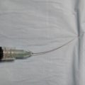
The senior author uses the FDA-cleared device, Selphyl (Aesthetic Factors, LLC, Wayne, NJ, USA) to produce an autologous PRFM. Peripheral blood is drawn from the patient into a vacuum collection tube containing a thixotropic separator gel. This tube is centrifuged for 6 minutes at 1100 rpm, which yields a supernatant plasma/platelet suspension and the cellular components (erythrocytes and leukocytes) below the separator gel ( Fig. 1 ). The plasma/platelet suspension is transferred to a second vacuum tube containing calcium chloride, which initiates the polymerization of fibrin. This polymerization process is completed in about 10 to 12 minutes ( Fig. 2 ) and the platelet-rich fibrin matrix can be injected through a 30-gauge needle before full polymerization. These platelets, embedded in the fibrin matrix, are capable of sustained release of PDGF, VEGF, TGF-B, and IGF-1 over 7 days.
In a clinical study, Sclafani showed that a single injection of PRFM below deep nasolabial folds could improve most moderate to deep nasolabial folds. The improvement was statistically significant within 14 days of treatment, and was stable for the remainder of the 3-month study. Sclafani later reported on his clinical experience with PRFM for facial uses, finding PRFM to be efficacious and well tolerated. Most patients required multiple treatments for optimal effect.
More recently, Sclafani and McCormick reported on the histologic changes associated with injection of PRFM into the dermis and subdermis in human skin. These investigators found significant new collagen deposition as early as 7 days, and significant angioneogenesis and adipogenesis clearly present by 19 days, without any evidence of cellular atypia.
Related posts:
 Latest Chemical Peel Innovations
Combining Fractional Carbon-Dioxide Laser Resurfacing with Face-Lift Surgery
Latest Chemical Peel Innovations
Combining Fractional Carbon-Dioxide Laser Resurfacing with Face-Lift Surgery
 Cannulas for Facial Filler Placement
Latest Innovations for Tattoo and Permanent Makeup Removal
Cannulas for Facial Filler Placement
Latest Innovations for Tattoo and Permanent Makeup Removal
 Cannulas for Facial Filler Placement
Stay updated, free articles. Join our Telegram channel

Full access? Get Clinical Tree


