Dermoscopy (dermatoscopy or surface microscopy) is an ancillary dermatologic tool that in experienced hands can improve the accuracy of diagnosis of a variety of benign and malignant pigmented skin tumors. The early and more accurate diagnosis of nonpigmented, or pink, tumors can also be assisted by dermoscopy. This review focuses on the dermoscopic diagnosis of pink lesions, with emphasis on blood vessel morphology and pattern. A 3-step algorithm is presented, which facilitates the timely and more accurate diagnosis of pink tumors and subsequently guides the management for such lesions.
Key points
- •
Dermoscopy is a valuable tool for the diagnosis of pink or nonpigmented skin lesions. Patient history and clinical examination, however, remain fundamental in helping to reach a correct diagnosis for a given lesion and represent the first step in deciding whether a particular lesion is tumoral (and possibly malignant) or whether it is part of an inflammatory or infectious process.
- •
Pink lesions lack pigment or are only partially pigmented. Therefore, the dermoscopic diagnosis of a given pink lesion relies on evaluation of the blood vessel types and patterns observed within it as well as on additional dermoscopic criteria, such as ulceration or scale. A 3-step algorithm has been formulated to assist in this regard.
- •
Benign pink lesions are typified dermoscopically by symmetry of color and pattern. Vessels seen on dermoscopy are usually of a single (or predominant) morphology and occur in a fairly regular pattern.
- •
Malignant pink tumors typically have a history of incessant growth. They often present on dermoscopy with an atypical or polymorphous vascular pattern (defined as having 2 or more vessel morphologies), with the vessels arranged irregularly. Additional clues for malignancy can also frequently be detected by dermoscopy.
- •
Dermoscopy has the potential to improve the detection of malignant pink tumors while reducing the number of excisions of benign pink lesions, for the ultimate benefit of patients.
Introduction
Pink, or nonpigmented, cutaneous lesions are a large heterogeneous group comprising both tumoral and inflammatory and infectious conditions. Pink tumoral (ie, neoplastic) lesions are the focus of this article. Nonpigmented skin tumors can present as macules, plaques, papules, or nodules and may be benign or malignant ( Table 1 ). Although clinical evaluation is fundamental for the diagnosis and subsequent management of these conditions, a naked eye diagnosis in many cases is inconclusive and dermoscopy has become a valuable ancillary diagnostic tool.
| Benign | Malignant | |
|---|---|---|
| Melanocytic | Compound and dermal nevi Red Clark nevi Spitz nevi | Primary AHM, including eczema-like melanoma CMMs |
| Nonmelanocytic | SH SK CCA Acantholytic dyskeratoma Angioma PG Dermatofibroma Benign adnexal lesions | BCC AKs SCCIS (Bowen disease) Invasive SCC Keratoacanthoma AS MCC Malignant adnexal lesions |
Pigmented structures within skin tumors provide important clues to improve the diagnosis of these lesions. Pink lesions lack or have scarce pigmentation, so in these cases the morphology and patterns of their blood vessels become essential in helping clinicians formulate a diagnosis (or differential diagnoses) and management plan. The definitions and morphology of these various vessels are provided in Table 2 . Table 3 details some important architectural or distribution patterns of vessels seen in pink tumors.
As shown in Table 4 , a 3-step algorithm can be used to help diagnose pink tumors. Initially, patient history and the overall clinical presentation of the lesion (or lesions) should be assessed to determine whether the latter is part of an inflammatory or infectious disease (such as psoriasis, lichen planus, scabies, or molluscum contagiosum) or whether it is tumoral. Once it is decided that the lesion (or lesions) is tumoral, the first step of the algorithm can be performed. In this step, the morphology of the vessels within the tumor is assessed. The second step involves noting the architectural pattern or distribution (arrangement) of the vessels within the lesion. The third step is to observe any additional dermoscopic features, such as remnants of pigmentation, scale, or ulceration. After performing these 3 steps, a diagnosis (or differential diagnoses) is formulated and an appropriate management plan organized.
| 1st Step: Vessel Morpholoy | 2nd Step: Vessel Arrangement | 3rd Step: Additional Features | Diagnosis | Management |
|---|---|---|---|---|
| Arborizing (branching) | Large-stem vessels, branching over lesion irregularly | Blue-gray ovoid nests and/or globules | nBCC or nodulocystic BCC | Biopsy |
| Fine microarborizing vessels, scattered irregularly | Multiple erosions; brown-gray leaflike and/or spoke-wheel areas, hublike pigmentation | sBCC | Biopsy | |
| Comma | Regular | Residual brown globules (or pseudonetwork on face), hairs | Congenital melanocytic nevus, compound or dermal nevus | No action |
| Dotted + comma | Regular | Pinkish-tan background pigmentation | Red Clark nevus | Follow up if similar to other nevi. Excise if solitary lesion. |
| Dotted | Regular | Reticular depigmentation; whitish striae; remnants of pigmentation (eg, brown globules, black dots, blue color) | Spitz nevus or thin AHM | Excision |
| Dotted | Stringlike (or reticular) | White halo or whitish background | CCA (or LCA) | No action |
| Dotted + glomerular | Clustered | Surface scale, white halo around vessels. | Bowen disease (SCCIS) | Biopsy |
| Dotted | Usually regular (central position) | Central whitish patch; delicate peripheral pigment network | DF | No action |
| Hairpin | Regular | Milia-like cysts; crypts; white halo around vessels | SK | No action |
| Radial or irregular (±polymorphous vessels) | White haloes and/or whitish background; central scale or keratin mass; ulceration/blood crusts; targetoid follicles; whitish pearls | SCC or KA | Excision | |
| Linear irregular | Irregular | Red homogeneous areas; pigment remnants (including blue-black color); ±hairpin vessels | Nodular AHM (or PG) | Excision |
| Linear irregular + dotted | Irregular | Red homogeneous areas; intersecting white rail lines; white collarette; ulceration | PG (or nodular AHM) | Excision |
| Central or irregular | Whitish striae; remnants of pink to brown-gray pigmentation | Thin or intermediate-thickness AHM | Excision | |
| Linear irregular + hairpin, corkscrew or arborizing | Central or irregular | Multiple colors; milky red globules or areas | Thick AHM or melanoma metastases | Excision |
| Red homogeneous areas | Throughout lesion | Intersecting white rail lines; white collarette; ulceration; ±linear irregular, dotted, hairpin vessels. | PG (or nodular AHM) | Excision |
| Crown | Radial | Central white to yellow lobular structures; ostia | SH | No action |
| Reddish pseudonetwork (face) | Confluent erythema located around hair follicles | Surface scale; targetoid follicles; linear-wavy vessels around follicles | AK | Topical therapy |
| Well-demarcated reddish lacunes | Regular | Whitish septae between lacunes | Angioma | No action |
Instrumentation and technique of dermoscopy
Regarding dermoscopic instrumentation, various handheld devices are available, which use either nonpolarized (eg, Heine Delta 20) or polarized light (eg, DermLite II Pro HR, DermLite III, or DermLite FOTO). Each modality has its strengths and limitations. For example, red color (eg, of erythema or vessels) and whitish streaks are enhanced visually with polarized instruments, whereas the white of milia cysts and blue and gray hues (eg, peppering seen in regression) are visualized better with nonpolarized light dermoscopy.
Generally, tumoral vessels are visualized well with noncontact polarized dermoscopy. A scaly surface atop a lesion may, however, obstruct the view of underlying vessels and make diagnosis difficult. In these situations, applying a fluid (eg, alcohol or immersion oil) onto the surface of the tumor helps reduce surface reflection from the scale, improving the visualization of underlying vascular features. Conversely, if the presence of surface scale is being assessed (eg, in cases of actinic keratosis [AK] or squamous cell carcinoma in situ [SCCIS]), dry dermoscopy without immersion fluid should be performed (using either a polarized or nonpolarized instrument).
Immersion fluids are routinely used in polarized or nonpolarized contact dermoscopy. In contact dermoscopy, care must be taken to exert minimal downward pressure on the tumoral surface. Excessive pressure can diminish or completely conceal vascular features and lead to difficulty with dermoscopic diagnosis. Immersion fluids (eg, oil) can be used in this context, but ultrasound gel is of higher viscosity and generally achieves superior results, that is, it permits good optical contact between the surface of the tumor and the glass plate of the dermatoscope even when the latter is applied lightly to the tumoral surface.
Benign pink (nonpigmented) lesions
Dermal Nevi
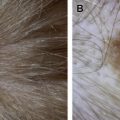
Dermal nevi are commonly occurring, benign melanocytic tumors that mostly develop in adulthood. They can be categorized into 2 main subtypes, which differ clinically and dermoscopically: Miescher nevi (on the face) and Unna nevi (located particularly on the trunk, extremities, and neck). Clinically, both Miescher and Unna nevi are usually long-standing lesions without a history of recent change. Miescher nevi, however, are smooth and dome-shaped, with a semifirm consistency (which may at times mimic nodular basal cell carcinoma [nBCC]). Conversely, Unna nevi are soft and papillomatous.
On dermoscopy, dermal nevi display a fairly regular arrangement of comma vessels. Remnants of pigmentation and hair may additionally be seen.
Both types of dermal nevi are characterized dermoscopically by coarse comma-shaped vessels, which are pinkish in color and slightly blurred. Comma vessels have a positive predictive value (PPV) for dermal nevi of 94% and are a significant negative predictor for amelanotic/hypomelanotic melanoma (AHM). The vessels of Miescher nevi are basically comma-shaped but can be somewhat variable in morphology (ie, varying in size and shape and frequently elongated) ( Fig. 1 A ). These vessels differ from the sharply focused, bright red arborizing telangiectasias of nBCC, which at times may be mistaken clinically for dermal nevus on the face. Like Miescher nevi, the comma vessels of Unna nevi are pinkish and slightly unfocused, but they differ in having a more classically comma shape, with less variation in size or shape (see Fig. 1 B).
Additional dermoscopic features of Miescher nevi include structureless tan-brown remnants of pigmentation and/or remnants of a pigmented pseudonetwork. Conversely, Unna nevi often display exophytic papillary structures, keratin-filled crypts, residual brown globules, and hairs.
Nonpigmented (Red) Clark Nevi
Nonpigmented or red Clark (atypical) nevi usually present as pink to pinkish-tan lesions of diameter greater than 5 mm in fair-skinned white patients (ie, skin phototype I or II). Red nevi are often multiple but may present as solitary lesions (ie, with or without the presence of other, pigmented nevi). Red nevi may present as macules, papules, or maculopapular tumors and can be difficult at times to distinguish clinically from AHM, particularly when presenting as ugly duckling lesions. Such ugly duckling lesions include solitary or relatively large red nevi.
Nonpigmented or red Clark nevi show a combination of dotted and comma vessels in a fairly regular arrangement, occurring on a pinkish-tan background. Comparison with other clinically similar lesions in patients should be performed to ensure comparable dermoscopic features of these nevi.
On dermoscopy, red Clark nevi typically show dotted and comma vessels, arranged fairly regularly throughout the lesions and occurring on a pinkish-tan background ( Fig. 2 ). The tan background may have a reticular or homogeneous appearance. These features generally contrast with nonpigmented Spitz nevi, which are usually solitary and fast growing and typically display a more dense arrangement of dot vessels on a pink-to-red background. In addition, reticular depigmentation, whitish striae, and remnants of pigmentation (eg, black dots, brown globules, and blue color) may additionally be seen in Spitz nevi. Dermoscopic examination of red nevi is also helpful in differentiating these tumors from AHM. Like Spitz nevi, AHM can present as solitary, fast-growing lesions and can exhibit a fairly dense arrangement of dot vessels on a pink-to-red background. In AHM, reticular depigmentation, whitish striae, remnants of pigmentation (eg, black dots, brown globules, blue color, and other melanoma-specific features), atypical vessels (eg, hairpin and linear irregular vessels) and ulceration may be additional clues for the diagnosis.
In patients presenting with multiple red Clark nevi, dermoscopic comparison of all the lesions should be performed to ensure comparable dermoscopic features of these nevi. To avoid missing a possible AHM, dermoscopic ugly duckling red Clark nevi–like tumors (ie, dermoscopically unlike the other red nevi on a given patient), solitary red tumors, lesions with a history of recent change, or lesions showing melanoma specific criteria should be excised.
Spitz Nevi
Spitz lesions were originally described in 1948 as juvenile melanomas by the pathologist, Sophie Spitz. Spitz believed that these lesions were childhood melanomas with a largely innocuous course (ie, rarely metastasizing, unlike adult melanomas). Of Spitz’ original 13 cases, at least 1 proved to be an actual malignancy (as evidenced by metastases and death), with the remainder having a benign biologic course.
On dermoscopy, flat nonpigmented Spitz nevi display regularly distributed dotted vessels on a pink to red background. Other features include reticular depigmentation, whitish striae, and pigment remnants.
Nonpigmented atypical or nodular Spitz tumors often show a polymorphous vascular pattern on dermoscopy.
Nonpigmented or hypopigmented Spitz lesions can closely mimic melanoma clinically and dermoscopically.
Spitz lesions nowadays are generally considered benign entities (ie, nevi) that can mimic malignant melanoma both clinically and histopathologically. Recently, however, some investigators have challenged this mainstream view and echoed Spitz’ original conclusions, hypothesizing that Spitz tumors may actually be a low-grade cutaneous malignancy with a high tendency to self-limitation and involution.
Spitz nevi classically present as fast-growing, smooth, dome-shaped papules or nodules. They are frequently pink or reddish in color but also commonly pigmented. They occur more commonly in children, especially on facial sites. As discussed previously, Spitz nevi assume significance because they are at times difficult to distinguish from melanoma on clinical grounds. In this setting, dermoscopy can be of assistance to clarify the diagnosis or at least raise a red flag for excision.
In flat nonpigmented Spitz nevi, dermoscopy stereotypically reveals dotted vessels that appear fairly closely aligned in a regular arrangement. The vessels lack whitish haloes and typically occur on a pink to red (milky red) background ( Fig. 3 A ). Dot vessels lacking white haloes are highly predictive for melanocytic skin tumors (PPV of 90%) and are particularly prevalent in Spitz nevi. Other dermoscopic features of flat Spitz nevi include reticular depigmentation (or negative network), a white network-like structure formed by intersecting white lines that surround the abovementioned dot vessels. White striae (chrysalis structures) can also be seen in some lesions and consist of shiny white orthogonal lines visible under polarized light dermoscopy. In Spitz nevi having pigment remnants, black dots, brown globules, bluish areas, and regions of pigmented network or streaks (ie, a residual starburst pattern) may also be apparent.
Nonpigmented atypical and nodular Spitz tumors typically display a polymorphous vascular pattern, with linear irregular vessels, coiled vessels, and/or milky red or pink globules. If partially pigmented, pigment remnants, such as homogeneous bluish areas and brown globules, may be visualized (see Fig. 3 B).
Because AHM, Spitz nevi, and atypical Spitz tumors can show similar dermoscopic features, excision should be performed for all nonpigmented spitzoid-looking lesions in adult patients. In children (ie, up to the age of 12 years), a more conservative approach is currently used when dealing with nonpigmented Spitz lesions exhibiting a stereotypical appearance. Contrarily, excision should be performed for large (>1 cm), nodular, rapidly changing, ulcerated, or otherwise atypical Spitz tumors appearing in childhood.
Sebaceous Hyperplasia
SH is a benign, nonpigmented neoplasm frequently presenting as multiple papules on the forehead, nose, and cheeks of middle-aged to elderly patients. Lesions are smooth, have a yellowish hue, and may have a central dell visible to the naked eye. Although a diagnosis is reliably made in many instances on clinical grounds alone, SH may at times be difficult to distinguish from other conditions, such as basal cell carcinoma (BCC).
Sebaceous hyperplasia (SH) is typified dermoscopically by crown vessels around the periphery of the lesion, with a white polylobular center.
Dermoscopy aids in the correct identification of SH by revealing crown (wreathlike) vessels, seen as pinkish, elongated, slightly blurred, and scarcely branching telangiectasias that surround the tumor (ie, in a radial arrangement). These vessels do not usually cross over the central parts of the lesion. The latter is in contrast to the arborizing telangiectasias of nBCC, which are typically bright red in color, sharply focused, and often pass over the center of the lesion. Multiple white to yellow, aggregated, homogeneous globular areas are also characteristically seen, which correspond histopathologically to the enlarged sebaceous lobules of SH. Moreover, a small centrally located yellowish crater (or craters) may be present on dermoscopy, which correlates with the dilated ostium (or ostia) of the infundibulum of the sebaceous glands ( Fig. 4 A ).
Seborrheic Keratosis
SKs are common benign acanthomas that commonly present as rough, well-demarcated, skin-colored to brown to black papules and plaques in adult patients. They are particularly prevalent on the trunk, face, and extremities and stereotypically have a stuck-on appearance. When nonpigmented, SK may clinically resemble other lesions, such as verruca, squamous cell carcinoma (SCC), or even AHM.
Nonpigmented seborrheic keratoses (SKs) display regularly distributed hairpin vessels, surrounded by a white halo. In addition, multiple milia cysts and crypts are often seen.
Dermoscopy assists in improving the accuracy of diagnosis of nonpigmented SK by revealing hairpin vessels (capillary loops) of fairly monomorphous morphology, arranged in a regular pattern. Hairpin vessels may occur in several different types of skin tumors but, when surrounded by white haloes, are suggestive of keratinizing lesions. Furthermore, hairpin vessels are particularly characteristic of SK, having a PPV for SK of 70%, compared with only approximately 13% for (invasive) SCC. In further contrast to SK, hairpin vessels in invasive SCC are usually elongated and irregular in morphology and occur in an irregular distribution.
Additional dermoscopic features of SK include a well-demarcated edge, a brainlike appearance with gyri and sulci (ridges and fissures), multiple whitish milia cysts, and pseudocomedone openings (crypts) (see Fig. 4 B).
Irritated or traumatized SK may at times pose a diagnostic and management dilemma, because they can clinically simulate malignant conditions, such as melanoma or SCC. This is especially the case if a history of recent trauma is not recalled by a patient. On dermoscopy, the hairpin vessels may be enlarged or elongated and appear somewhat irregular in morphology. Signs of recent trauma, namely blood crusts and/or erythema (inflammation), are also typically seen. Such features can make a dermoscopic diagnosis challenging. Remnant areas showing specific features of SK (ie, well-demarcated edge, multiple large milia cysts, crypts, fissures and ridges), however, may be present, which suggest the correct diagnosis of traumatized SK. Close clinical follow-up typically reveals that the features of trauma subside within a few weeks. In doubtful cases, however, where a diagnosis of malignancy cannot be ruled out, biopsy at the outset is recommended.
Benign pink lichenoid keratosis (LK) is considered a variant of SK and presents as a scaly or smooth, pinkish macule, plaque, or papule that may be pruritic. The lesion is especially prevalent on the trunk and extremities of middle-aged to elderly patients and can clinically mimic several lesions, such as psoriasis, AK, SCCIS, BCC, and even amelanotic melanoma (AM). Dermoscopically, surface scale may be highlighted and dotted or coiled and/or telangiectatic vessels may be seen, the vessels typically arranged in a fairly regular pattern ( Fig. 5 ). In lesions with pigment remnants, tan-gray blotches may be present. Because the dermoscopic features of pink LK can overlap with nonpigmented malignant tumors, however, such as early SCCIS, BCC, or AHM, biopsy is recommended.
Clear Cell Acanthoma (and Large Cell Acanthoma)
CCA, also called Degos or pale cell acanthoma, is an uncommon benign epithelial tumor that usually presents clinically as a solitary, pink to red plaque, papule, or nodule in middle-aged or elderly patients. Additionally, a scaly surface is sometimes present and a scaly collarette may be visible around the edge of the lesion. Many other benign and malignant nonpigmented skin lesions may resemble CCA clinically, including SK, BCC, SCC, and AM.
Clear cell acanthoma (CCA) reveals dotted and/or coiled vessels in a stringlike arrangement. A scaly surface and collarette may also be seen. Stringlike vessels may also occur in large cell acanthoma (LCA).
Dermoscopy assists in the identification of CCA by showing a striking pattern of dot and/or coiled vessels arranged in a linear, stringlike distribution ( Fig. 6 A ). At times, these linear patterns may assume a reticular arrangement. In addition, a whitish scaly surface may be highlighted by dry dermoscopy (ie, without immersion fluid) as well as a translucent to whitish peripheral scaly collarette. In contrast to the dot vessels seen in melanocytic lesions (including melanoma), the vessels in CCA are surrounded by whitish haloes or occur on a whitish background, as is customary for keratinizing tumors. Furthermore, the stringlike pattern of CCA contrasts with the typically regular arrangement of dot vessels in melanocytic skin lesions. In thicker variants of CCA, the vessels may appear coiled (or even glomerular), but they essentially retain their typical stringlike arrangement.
LCA is another rare, benign acanthoma that can at times present as a (scaly) pink papule or plaque on the trunk or extremities of middle-aged to elderly patients. Differential diagnoses may include SK, SCCIS (Bowen disease), BCC, and AM. Although considered highly specific for CCA, it is the experience of the authors that a stringlike (or reticular) arrangement of dotted and coiled vessels may also be seen in LCA (see Fig. 6 B). Further studies on the dermoscopic features of LCA, however, are needed.
Acantholytic Dyskeratotic Acanthoma (Acantholytic Dyskeratoma)
Acantholytic dyskeratotic acanthoma (acantholytic dyskeratoma) is a benign lesion typically presenting as a smooth to scaly-topped papular, pinkish tumor on the trunk of middle-aged to elderly patients. The lesion usually presents as a solitary tumor less than 1 cm in diameter and clinical differential diagnoses may include SK, verruca, nevus, BCC, AK, SCCIS, and invasive SCC.
Brown colored, scaly, starlike shapes can occur in acantholytic dyskeratoma, surrounded by a white to pink to tan background. Very fine and regular vessels may also be visible.
On dermoscopy, acantholytic dyskeratoma may reveal tan to darker brown–colored, starlike (stellate) shapes consisting of surface scale and/or superficial indentations (microfissures). These features are hardly visible with the unaided eye. Tan-brown areas can appear in some lesions as broken-up and branching lines and/or discrete rounded areas. Similar brown stellate shapes can be seen in papules of transient acantholytic dermatosis (Grover disease), another condition displaying acantholytic dyskeratosis on histopathology.
In addition, the tan-brown superficial scaly starlike areas typically have whitish edges and occur on a white to pink to tan background ( Fig. 7 ). Very faint and fine dotted hairpin and linear vessels may also be visible, having a regular morphology and whitish haloes, the latter feature typically seen in keratinizing tumors. The tan-brownish keratotic areas of acantholytic dyskeratoma may at times be relatively large and round or oval in shape, reminiscent dermoscopically of the central yellowish-brown keratotic plugs or (giant) pseudocomedones seen in Darier disease.
Related posts:

Stay updated, free articles. Join our Telegram channel

Full access? Get Clinical Tree


