, Shimin Chang2, Jian Lin3 and Dajiang Song1
(1)
Department of Orthopedic Surgery, Changzheng Hospital Second Military Medical University, Shanghai, China
(2)
Department of Orthopedic Surgery, Yangpu Hospital Tongji University School of Medicine, Shanghai, China
(3)
Department of Microsurgery, Xinhu Hospital Shanghai Jiao Tong University, Shanghai, China
Cutaneous branches from the thoracodorsal and lateral thoracic arteries were first mentioned by de Coninck et al., and the flaps based on these arteries have been known as thoracodorsal skin flaps, thoracodorsal axillary flaps, and lateral thoracic flaps [1–5].
17.1 Vascular Anatomy
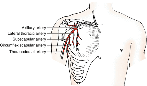
The subscapular artery arises from the axillary artery and divides into the thoracodorsal and the circumflex scapular arteries (Fig. 17.1). The thoracodorsal artery penetrates the latissimus dorsi muscle about 8–14 cm from the bifurcation of the subscapular artery into the circumflex scapular and thoracodorsal. Shortly before it enters the muscle, the vascular bundles send a branch to the serratus anterior muscle. The latissimus dorsi muscle is nourished by two main muscular branches from the thoracodorsal artery: a lateral branch running parallel to the anterior border and a horizontal branch passing obliquely to the dorsal and medial part of the muscle. They give multiple terminals to the skin as perforators through the muscle along the course of branches, and these musculocutaneous perforators are located at intervals in the back area. Septocutaneous perforators arise from the branch to the serratus anterior or other cutaneous branches, and they reach the skin between the latissimus dorsi and serratus anterior muscles.


Fig. 17.1
Vascular anatomy of the thoracodorsal and lateral thoracic artery
Cutaneous branches originate from the main thoracodorsal artery or from the branch to the serratus anterior.
They also supply septocutaneous perforators or direct cutaneous perforators.
17.2 Illustrative Case
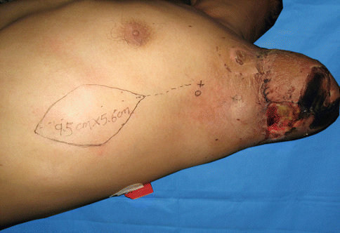
A 30-year-old male presents with complete left upper extremity amputation at proximal humerus after industrial grinder accident. The patient was placed in a lateral decubitus position with the ipsilateral upper extremity positioned freely and exposing the axilla by using adequate supports and padding, taking great care to avoid compression of the axillary neurovascular structures. The anterior border of the latissimus dorsi muscle and the center of the axillary fossa are marked (Fig. 17.2).


Fig. 17.2
Flap design
Stay updated, free articles. Join our Telegram channel

Full access? Get Clinical Tree


