, Shimin Chang2, Jian Lin3 and Dajiang Song1
(1)
Department of Orthopedic Surgery, Changzheng Hospital Second Military Medical University, Shanghai, China
(2)
Department of Orthopedic Surgery, Yangpu Hospital Tongji University School of Medicine, Shanghai, China
(3)
Department of Microsurgery, Xinhu Hospital Shanghai Jiao Tong University, Shanghai, China
Pedicled perforator flaps are also called local perforator flaps, or island perforator flaps. It combines the advantages of pedicled local flaps (good color and texture match, like-with-like reconstruction), pedicled regional flaps (up to 180° arc of rotation), pedicled distant flap (vascular reliable and larger size), and without microsurgical vascular anastomosis. For most small- to medium-sized defects, pedicled perforator flap allows linear closure of the donor site.
Theoretically, a flap can be designed and harvested based on any dominant and clinically relevant perforator. With over 300 perforators in the body, a large number of flaps can be theoretically harvested when an appropriate perforator is selected. The principle of free-style local perforator flaps can be used and applied to harvest pedicle perforator flaps for reconstruction of various head and neck, trunk, and extremity defects.
First of all, the perforators in the vicinity of the defect were localized by noninvasive methods, for example, Doppler, Duplex, angiography, or CTA, then the flap can be designed and elevated.
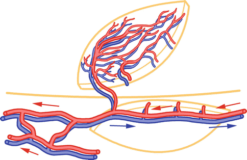
There are two types perforator-pedicled island flaps, V-Y advancement flaps and propeller flaps (Fig. 4.1). The distally pedicled version of perforator flaps is very useful for limb reparative and reconstructive surgery, especially for the distal extremities such as wrist-hand and foot-ankle region, which is the focused topic in this book.


Fig. 4.1
Propeller flaps. Arterial perfusion direction (red arrow), venous drainage direction (blue arrow)
4.1 Selection of Flap Movement Fashion
The most important factor to determine flap movement (advancement versus propeller) is the distance between the emerging point of the perforator and the proximal margin of the defect (perforator-defect distance). If an audible signal is recognized in Doppler only in close proximity to the defect (perforator-defect distance < defect diameter), the project of a V-Y advancement flap should be carefully evaluated, and a propeller flap movement should be chosen. On the other hand, if the perforators are detected at an intermediate distance from the defect (perforator-defect distance ≥ defect diameter), the application of the V-Y advancement model is recommended.
Generally speaking, V-Y advancement moves shorter distance than propeller rotation. V-Y advancement is more suitable for head-neck and trunk region and more suitable for myocutaneous perforators. Propeller movement is more suitable for limbs and septocutaneous perforators (Table 4.1).
Table 4.1
Comparison of perforator-pedicled V-Y advancement flaps and propeller flaps
V-Y advancement flaps | Propeller flaps | |
|---|---|---|
Pedicle movement | Advancement | Rotational |
Range of movement | Small-medium | Wide |
Flap size | Small-medium | Medium-large |
No. of perforators | Multiple | Single |
Possibility of converting to conventional “plan B” flap | Always maintained | Often maintained |
Vascular safety | High | High, but unpredictable |
Donor site | Primary closure | Skin graft for large defects |
4.2 Perforator-Pedicled V-Y Advancement Flaps
The perforator-based version of the V-Y flap, being based on a fully dissected and isolated perforator, has a range of movement that is considerably wider than a classic V-Y flap. Its advantages are simplicity, speed of harvesting, and donor-site closure. Its drawbacks are limited movement, limited size, and transfer over the defect of the skin lying in close proximity to the wound.
A V-Y advancement flap is indicated for small, noncomplicated wounds when a good perforator can be isolated. The amount of tissue that can be transferred to cover the defect is limited by the perforator caliber and the amount of advancement by the perforator length. The skeletonized perforator is very sensitive to stretching and shearing, and it must not be pulled too much. The flap must be accurately planned before the skin is incised circumferentially. To avoid wound dehiscence, it must reach the recipient site without any tension. In contrast, the donor-site appearance is excellent, and after a few weeks, the scars may be almost invisible. The morbidity of the donor site is minimal.
4.2.1 Flap Design and Dimension
The length of the flap is planned to be 1.5–2 times the diameter of the defect in the direction of advancement, while the width is slightly larger than the width of the defect. These suggestions about length-to-width ratio reflect the geometrical constitution of the V-Y advancement model. Adherence to these general rules rather than to the classic length-to-width ratio is considered clinically safe and recommended in practice, as it is with ease for wound closure after the skin island advancement. The perforator V-Y advancement flaps are ideally centered on the axial perforator vessel and do not act as random-pattern flaps.
4.2.2 Exploratory Incision and Pedicle Dissection
Flaps are harvested with the aid of 2.5× loupe magnification. The exploratory incision is carried down to the deep fascia. On the extremities, a subfascial approach is preferred to expose and dissect the perforators. Once all the perforators have been identified, their position in the flap, their caliber, their pulsatility, and the presence of adequate venae comitantes are evaluated. The perforators chosen to obtain an advancement movement are carefully dissected for 2–3 cm by gently teasing the muscular fibers. Perforators are irrigated intermittently with 2 % lidocaine solution during flap dissection. Extensive intramuscular dissection is not mandatory and is recommended only when pedicle elongation is needed to improve flap advancement.
4.2.3 Skin Paddle Circumcision
Stay updated, free articles. Join our Telegram channel

Full access? Get Clinical Tree


