This article outlines the epidemiology, pathogenesis, clinical presentation, diagnosis, and treatment of childhood morphea. Also known as localized scleroderma, morphea is a fibrosing disorder of the skin and subcutaneous tissues. Morphea is differentiated from systemic sclerosis (scleroderma) based on the absence of sclerodactyly, Raynaud phenomenon, and nail-fold capillary changes. Confusion may occur because patients with morphea often have systemic symptoms such as malaise, fatigue, arthralgias, myalgias, and positive autoantibodies. Unlike morphea, systemic sclerosis has organ involvement, particularly gastrointestinal, pulmonary, and renal.
Key points
- •
Morphea is a fibrosing disorder of the skin and subcutaneous tissues.
- •
The absence of sclerodactyly, Raynaud phenomenon, and nail-fold capillary changes distinguishes morphea from systemic sclerosis (scleroderma).
Background
Morphea, also known as localized scleroderma, is a fibrosing disorder of the skin and subcutaneous tissues. Morphea is differentiated from systemic sclerosis (scleroderma) based on the absence of sclerodactyly, Raynaud phenomenon, and nail-fold capillary changes. Confusion may occur because patients with morphea often have systemic symptoms such as malaise, fatigue, arthralgias, myalgias, and positive autoantibodies. Unlike morphea, systemic sclerosis has organ involvement, particularly gastrointestinal, pulmonary, and renal.
Epidemiology
The incidence of morphea is estimated at 0.4 to 2.7 per 100,000 population. A population-based study looked at morphea occurrence (adults and children) in Olmsted County, Minnesota, from 1960 to 1993, and estimated an annual incidence (age and sex adjusted) of 2.7 per 100,000 and a prevalence of 50 per 100,000.
Morphea is more common in whites and women, with a female to male ratio ranging from 2.4:1 to 4.2:1. Linear morphea, however, has an equal sex distribution. Morphea has a bimodal age presentation and is equally common in adults and children. Ninety percent of children present between 2 and 14 years of age. The mean age for adults presenting with morphea is in the mid-40s.
Plaque-type morphea is the most common type affecting adults ; linear morphea is most common in children. There is often a family history of other autoimmune diseases. Rarely there is a family history of morphea ( Fig. 1 ).
Pathogenesis
The pathogenesis of morphea is not fully understood. Imbalance of collagen production and destruction is thought to be an important factor. Inflammation and vascular changes are also important. Mononuclear cells infiltrate the skin and surrounding blood vessels in early morphea ; this leads to functional and structural changes to the microvascular system. Involvement is limited to tissues derived from the mesoderm.
Morphea plaques slowly soften over a period of 3 to 5 years when left untreated, but patients often develop new areas of involvement over their lifetime. Trauma and radiation have been reported before the onset of morphea. Morphea following vaccination has also been reported. A dental extraction has been reported before onset of ipsilateral progressive hemifacial atrophy.
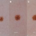
Elevated cytokines, soluble cell adhesion molecules, positive antinuclear antibodies (ANAs), and activated T lymphocytes have suggested autoimmunity as an important factor in the development of morphea. Certain medications, such as bisoprolol, bleomycin, peplomycin, d -penicillamine, bromocriptine, l -5-hydroxytryptophane in combination with carbidopa, pentazocine, and balicatib, have been associated with development of morphea. Borrelia has been implicated as a possible infectious cause of morphea, but other studies have refuted the association. Vascular injury has also been proposed as a cause of morphea, as supported by reduced capillaries and damaged endothelial cells. Long stretches of disease remission are common, but most patients with morphea will develop new lesions over their lifetime ( Fig. 2 ).
Clinical presentation
Peterson, in 1995, recommended the following classification system.
Five subgroups:
- •
Circumscribed (plaque) morphea
- •
Generalized morphea
- •
Bullous morphea
- •
Linear morphea (includes en coup de sabre and progressive hemifacial atrophy)
- •
Deep morphea (includes subcutaneous morphea, morphea profunda, disabling pansclerotic morphea, and eosinophilic fasciitis)
The classification system is controversial because it does not include the mixed types of morphea that can occur, and includes eosinophilic fasciitis as a subtype of morphea, which some clinicians consider a separate entity.
The initial inflammatory stage of morphea is characterized by erythematous or violaceous patches or plaques; the central region becomes white and sclerotic while the active borders remain red. Over time the active stage subsides, leaving behind sclerotic plaques that can be white or hyperpigmented. The overabundant collagen deposition destroys adnexal structures and hair follicles.
- •
Circumscribed morphea: fewer than 3 indurated discrete plaques ( Fig. 3 )
- ○
Can be superficial or deep
- ○
Deep variant (morphea profunda) affects the dermis and subcutaneous tissues
- ○
Can involve the underlying fascia and muscle
- ○
Often areas of pressure such as hips, waists, bra line
- ○
Breasts are commonly involved in women with sparing of the nipples
Fig. 3
Atrophic plaque on the buttock.
- ○
- •
Generalized morphea: greater than 4 indurated plaques larger than 3 cm and/or involving 2 or more body sites but sparing the face and hands ( Figs. 4–7 )
- ○
Rare variant, 7% to 9% of those affected with morphea
- ○
More likely to have positive autoantibodies, especially ANAs
- ○
Can have systemic symptoms such as myalgias, arthralgias, and fatigue
- ○
Often confused with scleroderma
- ○
Differentiated from systemic sclerosis by the absence of Raynaud phenomenon, nail-fold capillary changes, and sclerodactyly
- ○
Usually limited to the dermis, and rarely involves subcutaneous tissues
Fig. 4
Morphea on the arm of a child with generalized morphea. Note the indurated plaque next to an atrophic area.
Fig. 5
Atrophic areas on the face of a child with generalized morphea.
Fig. 6
Indurated plaques involving the hips and back in this child with generalized morphea.
Fig. 7
Indurated plaques involving the hips and back in this child with generalized morphea.
- ○
- •
Bullous morphea
- ○
Presents as tense subepidermal bullae that occur within affected sclerodermoid plaques
- ○
Rare variant, no known trigger
- ○
- •
Linear morphea ( Figs. 8 and 9 )
- ○
Most common type in children
- ○
Involves the lines of Blaschko, suggesting genetic mosaicism
- ○
Three variants include en coup de sabre, progressive hemifacial atrophy, and linear limb morphea
- ○
Associated with underlying tissue atrophy
- ○
En coup de sabre
- ▪
Usually occurs on the paramedian forehead
- ▪
Can be associated with underlying ocular and central nervous system (CNS) involvement, including headaches and seizures
- ▪
Typically follows Blaschko lines
- ▪
Can be associated with alopecia
- ▪
Can present (less commonly) with more than 1 lesion
- ▪
- ○
Progressive hemifacial atrophy (also known as Parry-Romberg syndrome) ( Fig. 10 )
- ▪
Minimal cutaneous changes with significant atrophy of the subcutaneous tissues
- ▪
Can have overlap with en coup de sabre; thought to be different ends of the same condition
- ▪
May have underlying seizures
- ▪
Mean age of onset of 13.6 years
Fig. 10
Progressive hemifacial atrophy.
- ▪
- ○
Linear limb morphea
- ▪
Associated with muscle atrophy, limb-length discrepancies, and joint contractures
- ▪
Usually unilateral
- ▪
- ○
Most likely to have extracutaneous manifestations with linear morphea
- ▪
One study looked at 750 children with linear morphea and found the following complications:
- •
Articular disease (47.2%), neurologic (17.1%), vascular (9.3%), ocular (8.3%), gastrointestinal (6.2%), respiratory (2.6%), cardiac (1%), and renal (1%)
- •
- ▪
Fig. 8
Linear morphea on the forehead of an adolescent male patient. Note the violaceous hue in this active plaque.
Fig. 9
Linea morphea in a female patient. Note the central white “burned-out” sclerotic area, with red advancing borders.Related posts:

Stay updated, free articles. Join our Telegram channel

Full access? Get Clinical Tree


- ○