Pectoralis Major Muscle and Musculocutaneous Flaps
S. ARIYAN
EDITORIAL COMMENT
This is one of the most significant musculocutaneous flaps for head and neck reconstruction. It is extremely versatile and has a hardy blood supply. The aesthetic appearance of the donor scar can be minimized by using the parasternal paddle, allowing for a curving midline scar with a transverse extension. This scar in women is almost completely hidden by the overlying breast.
INDICATIONS
The pectoralis major musculocutaneous flap can be used to reconstruct the oropharynx or the defect left after orbital exenteration or temporal bone resection as well as for mandibular reconstruction. (See Chapter 218 for reconstruction of the esophagus and Chapter 407 for chest wall reconstruction.)
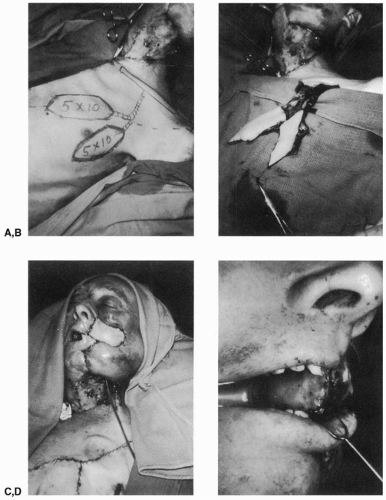
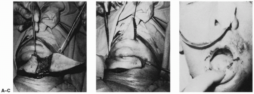
In most cases, tumors of the floor of the mouth or lateral tongue can be reconstructed by other flaps, such as the sternocleidomastoid, platysma, nasolabial, or tongue flaps. If the surgical wound is larger than these flaps can cover, however, more tissue can be provided with the pectoralis major musculocutaneous flap, with either a single skin paddle, two skin paddles in tandem (Fig. 135.1), or two split-skin paddles side by side (Fig. 135.2). Previously, reconstructions of the tongue and piriform sinus with forehead flaps or deltopectoral flaps did not provide sufficient bulk. These cutaneous flaps are relatively thin flaps, and reconstructions using them often led to a “funnel” that directed fluids toward the laryngeal airway, frequently leading to aspiration of fluids.
Resection of the lateral floor of the mouth, alveolar ridge, posterior half of the tongue, and piriform sinus requires sufficient skin and bulk for reconstruction. This can be achieved with a pectoralis major musculocutaneous flap in one stage. The bulk provided by this flap appears to be sufficient to avoid aspirations, either by directing fluids past the airway or by diverting fluids to the contralateral normal piriform sinus, as a result of fullness on the operated side.
Orbital Exenteration
The wound resulting from an orbital exenteration has been reconstructed by a variety of flaps, including forehead flaps (12) and temporalis muscle flaps (13, 14, 15, 16). The pectoralis major musculocutaneous flap is particularly useful for reconstruction in this area because it provides bulk and well-vascularized tissue to fill the cavity, to seal cerebrospinal fluid (CSF) leaks, and to provide greater protection against bacterial invasion (1, 2, 3).
Temporal Bone Resection
Resections of malignant tumors of the external auditory canal and mastoid can cause significant disability because of the proximity of the disease to the brain, the major dural sinuses, the carotid artery, and the cranial nerves, or because of the surgical treatment necessary to control the malignancy. Adequate tumor resection often has been compromised because of the complexity of the surgery as well as the difficulty of the reconstruction. In addition, these wounds often have been complicated by CSF leaks and bacterial contamination.
Because these cancers are otherwise uniformly fatal, treatment should consist of a radical en bloc resection of the tumor and temporal bone, together with a full course of postoperative radiation therapy (17, 18). Although these wounds have been covered with rotation scalp flaps and deltopectoral flaps, the pectoralis major musculocutaneous flap is ideal for these reconstructions because it provides sufficient bulk and soft tissue to cover the dura and seal it against CSF leaks and it provides a rich vascularity to permit uncomplicated healing despite bacterial contamination of the wounds (19).
Mandibular Reconstruction
Bone grafts often fail to reconstruct a mandibular segment because of the poor vascularity in the recipient bed resulting
from radiation fibrosis or dermal scarring from previous surgical procedures or injuries.
from radiation fibrosis or dermal scarring from previous surgical procedures or injuries.
 FIGURE 135.2 A: A split pectoralis major muscle with two side-by-side skin paddles. B,C: These were used to line and resurface a through-and-through defect of the cheek. D: The lining at 3 weeks. |
Because I was able to demonstrate that rib grafts would survive on their periosteal blood supply, I then employed the pectoralis major musculocutaneous flap, incorporating a segment of underlying rib to reconstruct a jaw (9). Although this extended pectoralis major flap may be used for reconstruction of segmental resections of the mandible, it does not appear to be suitable for reconstruction of the entire mandible.
ANATOMY
Although the thoracoacromial artery had been previously described in the literature as running along the undersurface of the pectoralis minor muscle, fresh cadaver dissections confirmed the consistent presence of this vessel along the undersurface of the pectoralis major muscle, with a branch from this vessel to the pectoralis minor. There are additional arteries to the pectoralis major, namely, the lateral thoracic artery to the lateral and inferior portions of the muscle, the superior thoracic artery to the clavicular portion, and the intercostal perforators from the internal mammary artery, providing some supply to the sternal portion of the muscle (2, 20, 21, 22).
Stay updated, free articles. Join our Telegram channel

Full access? Get Clinical Tree


