57. Liposuction
First recorded attempt at lipectomy is attributed to the French surgeon Dujarier, who, in the 1920s, attempted to remove fat from a dancer’s calves using a uterine curette. 1
Vascular damage resulted in amputation of the leg.
In the mid-1970s Giorgio Fischer and his father, Arpad Fischer, developed the “cellusuctiotome,” an instrument made of a hollow curette and blade attached to a suction pump. This method had a high rate of bleeding complications.
Yves-Gerard Illouz and Pierre Fournier improved on the prior techniques by replacing the sharp curettes of the 1970s with a cannula and suction system, the introduction of a “wetting solution” containing saline solution and hyaluronidase, and the use of a “crisscross” technique.
These methods decreased bleeding and contour-associated complications.
In the 1980s a dermatologist, Jeffrey Klein, introduced the tumescent technique.
Tumescent technique: Subcutaneous infiltration of a large volume of diluted lidocaine and epinephrine that expands the fat compartment causing it to become swollen and firm, or tumescent
Provides local anesthesia and reduces blood loss
Anatomy
Subcutaneous Layers
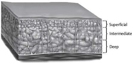
Subcutaneous adipose tissue is divided into superficial and deep layers throughout the body by Scarpa fascia or the superficial fascial equivalent. 2
For purposes of body contouring, the subcutaneous fat is arbitrarily divided into three layers. 3
Superficial
Dense fat, adherent to overlying skin
Aggressive, avulsive, or thermal liposuction methods should be used with great Caution in this layer to prevent contour irregularities and skin damage.
Intermediate
Safest layer
Most commonly suctioned layer
Deep
Loose and less compact layer
Can be removed safely in most areas
Zones of Adherence
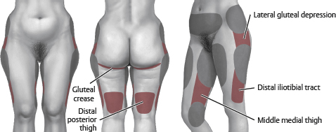
Distal iliotibial tract
Gluteal crease
Lateral gluteal depression
Middle medial thigh
Distal posterior thigh
Caution: Be very cautious removing fat from zones of adherence. The stiff fibrous network predisposes these areas to postoperative contour deformities.
Senior Author Tip:
The rigid fibrous connections at zones of adherence can be relaxed using an exploded tip cannula without suction (i.e., separation and equalization). The tissue will become more pliable to allow smooth transitions into surrounding areas and enable these zones to be traversed or treated safely
Cellulite (Gynoid Lipodystrophy)
Peau d’orange and mattresslike deformity seen primarily in women and obese patients
Two types 4
Primary or cellulite of adiposity: Results from hypertrophic fat cells in the superficial fat between the septa of the superficial fascial system
Typically present when supine and erect, seen in younger women
Generally not improved with skin-tightening procedures
Secondary or cellulite of laxity: Results from increased skin and superficial fascial system laxity
Present when erect but not supine, usually >35 years of age
Treated with skin- and superficial fascial system-tightening procedures
Preoperative evaluation
Physical Examination
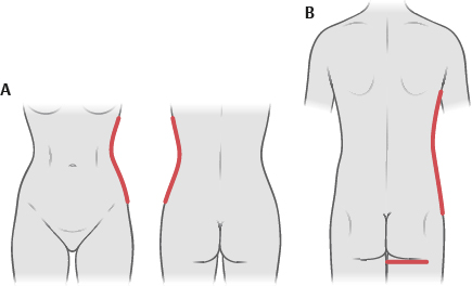
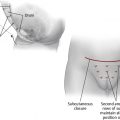
Check for deviation from ideal contour (Fig. 57-3).
Female ideal contour
Concavity below the rib cage that changes to a convexity over the hips and thighs
Medial and lateral thighs have mild convexities.
The buttock crease blends laterally with thigh.
Male ideal contour
More linear silhouette, less concavity and convexity below rib cage and over thighs
The buttock crease is squared and linear.
Flat anterior infraumbilical region
Note any of the following:
Asymmetries
Dimpling/cellulite
Location of fat deposits
Areas of adherence
Hernias and myofascial diastasis
Check skin laxity.
Examine spine for scoliosis.
May cause asymmetry
Assess for hernias/diastasis.
Medical History
Agents that interfere with coagulation should be avoided.
Aspirin
NSAIDs
St. John’s wort
Vitamin E
Herbal supplements
Other anticoagulants
Note personal and family history of deep venous thrombosis or clotting disorders.
Photographs
Standard photographs of areas to be treated should be obtained.
See Chapter 3 for further details on photography.
Senior Author Tip:
Patient selection is critical. Common pitfalls include: redundant and poor quality skin, obesity with excess intraabdominal fat and unreasonable expectations
Perioperative Considerations
Preoperative
Complete blood cell count if expecting to perform large-volume (>5 L total lipoaspirate) procedure
Perioperative IV antibiotics
Deep venous thrombosis prophylaxis
Intermittent pneumatic compression devices should be used intraoperatively.
Chemoprophylaxis may be given to those at higher risk (see Chapter 11).
Hypothermia
Forced-air warming blankets
Consider circulating warm water mattresses
Cover exposed body areas.
Warm intravenous fluids.
Warm operating room.
Warm wetting solutions.
Positioning
Pad all pressure points.
Prone position
Protect face, breasts, and genitals.
Soft hip roll beneath iliac crest
Supine position
Arm abduction <90 degrees to prevent brachial plexus injury
Hips and knees flexed at 30 degrees with a pillow
Senior Author Tip:
Using multiple patient positions allows for target areas to be treated thoroughly, without the distortion and compression from the operating table seen when using a single supine position, or even in supine/prone positioning. Three positions: Supine, lateral decubitus, and the opposite lateral decubitus allow for complete exposure of the body circumferentially while avoiding the more onerous and time-consuming prone position. As a caveat, if the operating surgeon is strongly one-handed, adding the prone position to the standard three position routine can help intraoperative assessment and prevent asymmetries. Additionally, multiple patient positions allow cross-hatching and help ensure complete treatment. It also reduces the risk of creating iatrogenic contour deformities
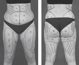
Markings
Patients should be marked when they are in an upright position or standing.
Use marker to outline areas to be treated.
Mark zones of adherence and other areas to be avoided with parallel lines or cross-hatch marks.
Incisions
Longer for ultrasound-assisted liposuction (UAL) compared with suction-assisted liposuction (SAL) (6-8 mm versus 2-3 mm, respectively).
Incisions can be placed anywhere adjacent to areas being treated.
Multiple incisions are used for access to target areas, and ideally they are strategically located to allow crisscross suctioning.
Liposuction from a single access incision may lead to contour deformity.
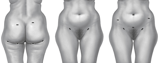
Box 57-1% Incision Locations for Liposuction
Breast (male): Anterior axillary fold and/or periareolar
Lateral back: Lateral bra line
Vertical back: Midline
Flank/hip: Sacral, groin crease, midaxillary line in panty line
Abdomen: Lateral lower abdomen/suprapubic/umbilical
Buttock: Sacral, midaxillary line in panty line
Lateral thigh: Midaxillary line in panty line
Posterior thigh: Midaxillary line in panty line
Medial thigh: Medial groin crease and inguinal crease
Anterior thigh: Inguinal crease
Upper arm: Anterior and posterior axillary folds, olecranon radial elbow crease
Liposuction Cannulas
Most tips are blunt with multiple openings set back from the end to allow suctioning of fat with passage of the cannula.
Blunt tips limit risks of penetration of unwanted structures such as fascia, peritoneum, vessels, and nerves.
Suction cannulas range from 1.8 mm up to 1 cm in diameter (typical use for liposuction is 2.5-5.0 mm) with varying cannula lengths.
Larger suction cannulas are typically used for deeper tissue.
As suction cannula size increases, the rate of fat removal with each pass increases, as does the risk of contour irregularities.
Physics and Theory of Liposuction
SAL removes fragmented fat through a cannula and tubing into a receptacle.
Fragmentation of fat
“Jackhammer effect”: The cannula striking fatty tissue
The avulsion of fat into the islets of the cannula as the cannula moves in and out
Rate of fat aspiration
Directly proportional to the diameter of the cannula and suction tubing
Directly proportional to vacuum pressure
Inversely proportional to the length of the cannula
Poiseuille law concepts
R = (L/r 4 ) × K, where R is the resistance, r is the radius of the tube, L is the length of the tube, and K is a constant factor
Wetting Solutions
Purposes
Volume replacement
Hemostasis
Analgesia
Enhance cavitation (UAL)
Dissipate heat
Constituents vary, examples:
1000 ml of lactated Ringer solution at 21° C
30 ml of 1% lidocaine plain (15 ml if large volume)
1 ml of 1:1000 epinephrine
Klein recipe 7
1000 ml normal saline solution
50 ml 1% lidocaine plain
1 ml 1:1000 epinephrine
12.5 ml of 8.4% sodium bicarbonate
Alkalization may decrease pain with infiltration, but is not needed with general anesthesia.
Lidocaine in Wetting Solution
Analgesia is provided for up to 18 hours postoperatively.
Recommended maximum is 7 mg/kg in the presence of epinephrine (4 mg/kg in the absence of epinephrine).
The estimated maximum safe lidocaine dosage using the tumescent technique is 35 mg/kg.
Peak plasma concentration is 10-14 hours after infiltration.
Klein’s original study Noted doses up to 52 mg/kg with no adverse effect; this has been confirmed in other studies.
Objective signs of lidocaine toxicity at plasma concentration >5 µg/ml
Use of high quantities of lidocaine made possible because of:
Diluted solution
Slow infiltration
Vasoconstriction of epinephrine
Relative avascularity of fatty layer
High lipid solubility of lidocaine
Compression of vessels by infiltrate
Note:
The wet environment may be lost after 20-30 minutes.
Related posts:

Stay updated, free articles. Join our Telegram channel

Full access? Get Clinical Tree


