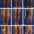
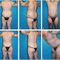
CHAPTER 28 Gluteal Liposculpting for Men
Summary
The chapter will allow the reader to understand the anatomy and the fundamental techniques for gluteal sculpting for men. From patient selection, to physical examination, to concise steps describing the technique, the reader will be able to have the basic tools necessary to create substantially improved results for male patients in their practice.
Introduction
While there are various techniques for contouring and augmentation of the buttock, fat grafting has become a preferred procedure for plastic surgeons. The use of fat for large-volume expansion, minor contour improvements, and restorative medicine has demonstrated the increased value placed on this body component. By providing a three-dimensional approach to reshaping, redistributing, and enhancing volume, fat grafting offers a precise, versatile method for buttock contouring, with a faster recovery and lower complication profile. Proponents of gluteal implants acknowledge that better patient and technique selection have reduced the immediate risks (e.g., wound dehiscence and prosthesis exposure, seroma, and infection) from 30% to 40% to a more acceptable 5%. Nevertheless, gluteal fat grafting altogether avoids the risks specific to implants, including capsular contracture, extrusion, and malrotation.
While gluteal liposculpting was initially popularized in the early 2000s, general fat- grafting techniques have evolved significantly over the past century. Fat grafting was first reported by Neuber in 1893. It was not until 1980, however, that the technique was truly pioneered by Illouz in Brazil, with Coleman pioneering techniques in long-term preservation of fat in vitro. Recently, grafting techniques have continued to evolve both as aesthetic and reconstructive procedures, with both academic institutions and biotechnology companies researching new applications as well as improving methods for fat harvesting, collection, preparation, and delivery. Key areas of focus include the long-term viability of grafts as well as developing a means for supercharging fat grafts with the addition of adipose stem cells. The major technical challenge involves recognizing whether there is sufficient fat available for harvest.
Facts about Fat
The number of fat cells is influenced by genetics, developmental periods, and eating habits. Infants are born with roughly 10 billion fat cells; as people age, healthy individuals might have 10 to 20 billion fat cells, compared to 100 billion fat cells in obese individuals.
Fat Production
While fat production is apparently genetically determined, humans can produce fat cells during the third trimester of pregnancy, at puberty, and in the setting of morbid obesity—all because of sex hormone production. Besides these conditions, fat cells will increase or decrease in size based on weight gain or loss, and density and number of cells remain the same. Therefore, the shape of an individual’s frame is genetically determined and can only be altered by removing or reshifting the location of fat cells through liposuction and/or fat grafting. This concept allows patients with large buttocks to benefit from gluteal liposculpting by making them smaller and shapelier despite the added volume.
Fat Availability Versus Survivability
There are many unknowns remaining in the pursuit of understanding the survivability of fat cells. Issues to address include viability of cells in lipoaspirate, optimization of donor and recipient sites, and the impact of endogenous and exogenous factors that may affect transfer.
A key distinction must be made between the amount of fat cells available for transfer versus the amount of fat cells that survive the transfer, with the ultimate goal to ensure the highest fat survival rate after transfer. While there are no in vivo methods of measuring cell viability or adipose-derived stem cell content, there are several in vitro measures, which currently lack clinical application. Nevertheless, there appear to be three types of cell death caused by particular mechanisms of injury:
Mechanical injury (e.g., suction).
Traumatic injury (e.g., blunt force).
Chemical injury (e.g., lidocaine toxicity).
Anatomy
Anatomical differences in the male patient are discussed in Chapter 30.
Anatomical Considerations: Deep and Superficial Fat Layers
The superficial fat layer varies in thickness from greater than or equal to that of the deep layer to as thin as 1/4 inch. In cases with a thick superficial layer with relative ease in fat extraction and no dermolipography, power-assisted liposuction (PAL) is used, sometimes in conjunction with Body-Jet (water-assisted liposuction). When discerning the deep from superficial layers is difficult, use of VASER (ultrasound- assisted liposuction [UAL]) or pretunneling with post-tunneling are helpful.
Patient Selection
The ideal candidate for gluteal liposculpting is slightly overweight but healthy and able to provide enough donor tissue for transfer. The more donor fat available, the more impressive the contour will be; therefore, larger patients can still be considered good candidates for the procedure. To determine preoperatively whether the patient has enough donor fat, a pinch test can estimate the amount available. The range of fat needed is between 450 and 1,100 cc per side, approximately. Patients must be American Society of Anesthesiologists class I or II only.
Steps for Gluteal Sculpting
Planning
Instruments and Equipment
Often a combination of liposuction technologies is used in the process:
PAL (nearly all cases).
UAL (60%–70%).
Body-Jet water–assisted liposuction (30%).
PAL is the workhorse technology. It provides an adjustable, controlled reciprocating cannula (4,000– 6,000 cycles per minute), increasing the speed of the procedure while reducing operator fatigue. The PAL system has multiple cannula tips and is best suited for fibrous fatty tissue, revision liposuction, and large-volume cases.
UAL technology functions by creating microbubbles that push apart fat tissue only while sparing all other tissue types, and thus loosen fatty tissue to be aspirated with the tumescent fluid in a process termed acoustic streaming. UAL is useful in areas of dense, fibrous fat, as well as in massive weight loss patients with “fluffy” fat, which can be particularly challenging to aspirate.
The VASER probe is a preferred UAL instrument, with different-sized probes creating different amplitude waves. The larger the probe cross-sectional area, the larger the amplitude and thus the greater the energy delivered. The 3.7-mm probe is suitable for most cases. While there are two probe modes, the VASER (pulse) mode is preferable in delicate areas, as it creates virtually the same effect with decreased energy delivery. The continuous mode is for typical use.
The Body-Jet system separates fat cells by water pressure using edematous disintegration, giving the fat cells a thinner, more liquid-like quality with removal. It causes the least amount of tissue trauma and ultimately less bruising.
Technique and the Influence of Fat Characteristics
Skin-to-Fat Relationship
When introducing the cannula, the creation of track marks on the skin surface indicates that the patient has a strong and direct association between fat and skin surface. This is termed dermolipography. Patients with a strong association have more compact layers of fatty tissue and require mechanical disruption via VASER ultrasound pulsed power or tunneling without suction, while those without marks can undergo suction extremely superficially.
Extraction Properties
In cases with light or fluffy fat, fatty tissue can be harvested rapidly and easily. In patients with more fibrous or resistant fat, the VASER at an 80% pulsed setting along with a sharper cannula can increase speed and efficiency of extraction.
Fat Cell Fragility
If an excess of oil is noted in the aspirate, the Body- Jet system provides a gentler extraction technique to prevent damage of more fragile fat cells.
Preoperative Preparation
Markings
Patients are photographed the morning of surgery with full 360° view with various angles, both with and without markings—the photographs are crucial for intraoperative use. Markings are made with the patient standing, identifying areas for liposuction. Zones 1 through 4 are always included, while zones 5 and 8 are often considered. Additionally, the posterior superior iliac spine is marked as the height of the gluteal muscle, making the V zone (zone 1) easier to identify and mark.
Medications
Patient are given 1 L of intravenous (IV) fluid preoperatively, along with methylprednisolone 125 mg IV and clindamycin 600 mg IV. Patients at high risk for postoperative nausea or vomiting receive ondansetron sublingually. Near the completion of the operation, dexamethasone 10 mg IV is given to reduce swelling and nausea in the postoperative period.
Related posts:
Stay updated, free articles. Join our Telegram channel

Full access? Get Clinical Tree


