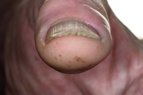
Paronychia


Paronychia


Richard B. Mallett


Evidence Levels:  A Double-blind study  B Clinical trial ≥ 20 subjects  C Clinical trial < 20 subjects  D Series ≥ 5 subjects  E Anecdotal case reports


image


Paronychia is characterized by inflammation of the proximal and/or lateral nailfolds, the fingers being more commonly affected than the toes. Acute paronychia is a painful pyogenic infection that usually occurs after injury or minor trauma and is characteristically caused by Staphylococcus aureus, although other aerobic and anaerobic bacteria have also been found.


Chronic paronychia, one of the commonest nail disorders, presents as tender erythema of the nailfolds with thickening of the tissues, loss of the cuticle, and subsequent dystrophy of the nail plate. The causative factors are repetitive microtrauma and exposure to water, irritants, and allergens, causing a contact dermatitis with subsequent colonization by yeasts and bacteria. Other less common causes of chronic paronychia include retronychia, characterized by the disruption of the longitudinal growth of a nail due to acute injury from physical or systemic causes, with resultant embedding of the old nail in the ventral surface of the proximal nailfold as the new nail regenerates. Also, cutaneous leishmaniasis may rarely present as an unusual chronic paronychia in endemic areas. Pemphigus vulgaris may present with either acute or chronic paronychia.


Drug-induced paronychia with pseudopyogenic granuloma is increasingly recognized and may occur with systemic retinoids, antiretroviral drugs such as indinavir, epidermal growth factor (EGF) receptor inhibitors, including gefitinib and cetuximab, and the novel anticancer mTOR inhibitors such as everolimus.


Tumors including Bowen’s disease, keratoacanthomas, squamous cell carcinoma, enchondroma, and amelanotic melanoma may masquerade as chronic paronychia.


Management strategy


Acute paronychia requires urgent effective treatment to prevent damage to the nail matrix. If the infection is superficial and pointing, then incision and drainage without anesthesia is possible. Infection is often due to S. aureus, but β-hemolytic streptococci and anaerobic organisms may also be found. A swab must be taken for bacterial culture and antibiotic sensitivity, and a broad-spectrum antibiotic covering both aerobic and anaerobic organisms given. Warm compresses with an astringent (e.g., aluminum acetate lotion, if available) can help reduce edema and provide a hostile environment for bacteria. For deeper infections, if there has been no marked clinical improvement after 48 hours of antibiotic therapy, surgical treatment should be undertaken. Under local anesthesia, the proximal third of the nail plate is removed and a gauze wick is laid under the proximal nailfold to allow drainage.


Chronic paronychia is usually due to dermatitis and often associated with wet work (e.g., in domestic workers, cooks, bartenders, fishmongers, etc.), and may be exacerbated by contact irritants or allergens. Immediate sensitivity to fresh foods can be a factor. In children, thumb sucking may initiate the condition. Eczema or psoriasis may predispose to chronic paronychia, as may poor peripheral circulation and rarely pemphigus vulgaris. Microtrauma, including over-zealous manicuring of the cuticle, is also important. The middle and index fingers of the right hand and the middle finger of the left hand are most commonly affected, but any finger may be involved. Inflammation with bolstering of the nailfold and loss of the cuticle opens a space between the nailfold and the nail plate, which commonly becomes infected with yeast, especially Candida species, and a wide range of other microorganisms. Acute exacerbations due to bacterial infection may occur.


Successful treatment relies on protection of the affected fingers from water, irritants, allergens, and trauma, together with anti-inflammatory treatment using moderately potent or potent topical corticosteroids. Tacrolimus 0.1% ointment applied twice daily may also be effective. Swabs for yeast and bacteria should be taken, anticandidal preparations can be useful, and antibiotic preparations may also be needed. Treatment should be continued until the inflammation has subsided and the cuticle reformed and reattached to the nail plate (3 months or more). Applying 80% phenol with a toothpick to the groove under the proximal nailfold may encourage reattachment. For frequent acute episodes, intralesional or systemic corticosteroids plus systemic antibiotics for a week may be useful. In cases where conservative management fails, surgery or low-dose superficial radiotherapy may be considered. For cases secondary to retronychia simple avulsion of the nail plate can be curative.


Drug-induced pseudopyogenic granulomatous paronychia responds to daily topical 2% mupirocin with clobetasol propionate ointment. Additionally, for patients on EGF or mTOR inhibitors oral doxycycline 100 mg bid or dose reduction may be useful.


Specific investigations


Stay updated, free articles. Join our Telegram channel

Aug 7, 2016 | Posted by in Dermatology | Comments Off on Paronychia

Full access? Get Clinical Tree

Get Clinical Tree app for offline access