Paediatric dermatoses


Paediatric dermatoses


Jane C Ravenscroft MRCP, MRCGP


Introduction

Skin conditions are very common in children. Many are transient and of little importance; however, some may have important implications. Atopic eczema is very common, and because of this, any rash in a child is often diagnosed as atopic eczema. This chapter will illustrate some of the many different skin lesions (Table 2.1) and rashes (Table 2.2) which can affect children, and help in accurate diagnosis and management.


Table 2.1 Lesions presenting in children <10 years, by colour, or presence of blisters


Red/pink/blue


• Abscess/boil


• Infantile haemangioma


• Lymphangioma circumscriptum


• Naevus flammeus


• Pilomatricoma


• Port wine stain


• Pyogenic granuloma


• Ringworm


• Spider naevus


• Spitz naevus


• Vascular malformations


Skin coloured/yellow/orange


• Aplasia cutis


• Dermoid cyst


• Epidermal cyst


• Granuloma annulare


• Molluscum contagiosum


• Neurofibroma


• Sebaceous naevus


• Xanthogranuloma


Pigmented


• Acquired benign naevus


Café au lait macule


• Congenital melanocytic naevus


• Dermatofibroma


• Epidermal naevus


• Mastocytoma


• Mongolian blue spot


• Naevus spilus


Hypopigmented


• Naevus depigmentosus


• Postinflammatory hypopigmentation


• Pityriasis alba


• Vitiligo


Blisters


• Bullous impetigo


• Chicken pox


• Chronic bullous disease of childhood


• Dermatitis herpetiformis


• Epidermolysis bullosa


• Herpes simplex


• Mastocytosis


• Miliaria


• Papular urticaria


Table 2.2 Rashes in children <10 years


Neonatal and infantile rashes


• Atopic eczema


• Epidermolysis bullosa


• Erythema toxicum neonatorum


• Ichthyosis


• Infantile acne


• Miliaria crystallina


• Miliaria rubra


• Psoriasis


• Seborrhoeic dermatitis


Rashes affecting the nappy area


• Atopic eczema


• Bullous impetigo


Candida infection


• Irritant nappy rash


• Langerhans cell histiocytosis


• Perianal streptococcal disease


• Psoriasis


• Seborrhoeic dermatitis


Acute childhood rashes


• Folliculitis


• Henoch-Schönlein purpura


• Infections:


– Chicken pox


– Erythema infectiosum


– Hand, foot, and mouth disease


– Herpes simplex


– Kawasaki disease


– Measles


– Roseola


– Rubella


– Scabies


– Scarlet fever


– Staphylococcal scalded skin syndrome


• Pityriasis rosea


• Toxic erythema


• Urticaria


Chronic childhood rashes


• Acne


• Atopic eczema


• Chronic bullous disease of childhood


• Dermatitis herpetiformis


• Ichthyosis


• Juvenile plantar dermatosis


• Keratosis pilaris


• Pityriasis versicolour


• Psoriasis


• Urticaria pigmentosum


SKIN LESIONS


Birthmarks


Infantile haemangioma (strawberry naevus)


Clinical presentation

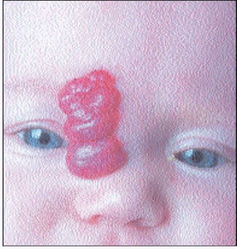
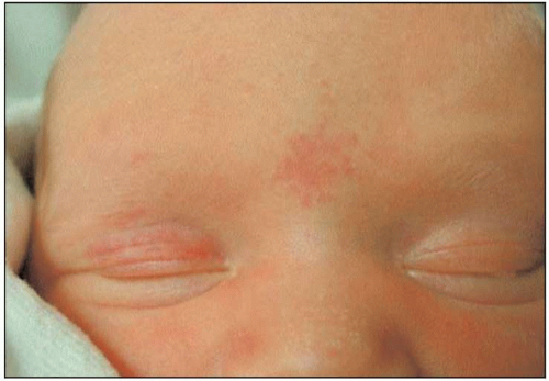
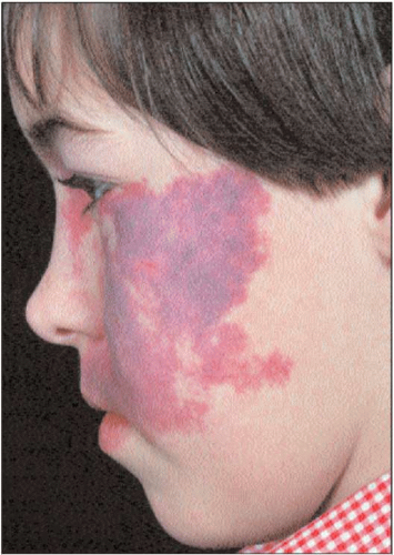
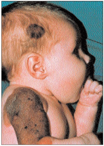
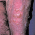
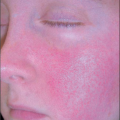
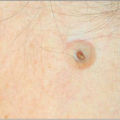
Infantile haemangiomas are benign vascular tumours which affect 1-2% of infants, most commonly on the head and neck. They are not present, or just visible at birth, and grow rapidly in the first 6 months of life to become distinctive bright red nodules, sometimes with a deeper component (
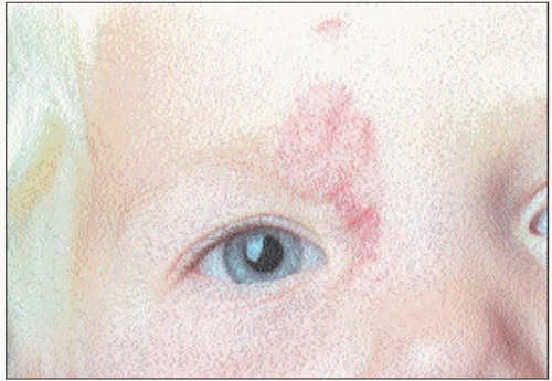
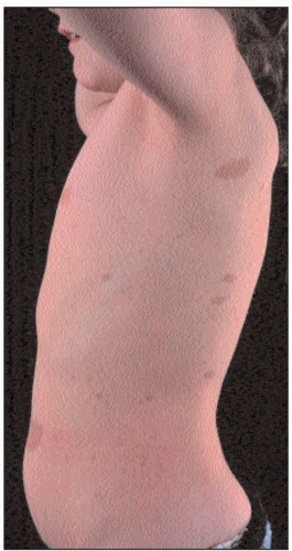
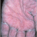
2.1). At around 12 months of age, they start to undergo spontaneous resolution (
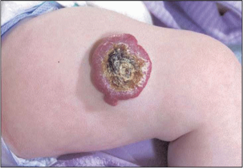
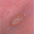
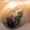
2.2). Fifty per cent of lesions will disappear by 5 years of age and 90% by 9 years. There may be residual flaccid skin and telangiectasia. Complications of ulceration (
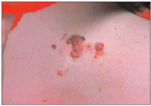
2.3), haemorrhage, and interference with function may occur during the phase of rapid proliferation. Rarely, multiple haemangiomas occur, which may be associated with internal haemangiomas. Very infrequently, large haemangiomas may be associated with structural brain and cardiac abnormalities, or consumptive coagulopathy.


Differential diagnosis

Differential diagnosis is from other vascular birthmarks. Vascular lesions which do not show characteristic proliferation after birth are not infantile haemangiomas and should be referred for accurate diagnosis.


Management

Due to the self-limiting nature of infantile haemangiomas, treatment is not required unless complications occur. Ulcerated lesions can be treated with topical steroid and antibiotics. Pulsed dye laser may be used for prolonged ulceration, and for residual telangiectasia after resolution. Interference with function may require intralesional or oral steroids.


Naevus flammeus (salmon patch)


Clinical presentation

This is a pale pink vascular patch, present at birth, which commonly occurs on the nape of the neck, forehead, or eyelid (2.4).


Differential diagnosis

Naevus flammeus can be distinguished from port wine stain by its paler colour and typical site.


2.1 Infantile haemangioma at 5 months of age.


2.2 The same child as in 2.1 aged 3 years.


2.3 A proliferating infantile haemangioma, which is ulcerated.


2.4 A salmon patch in a neonate.


Management

Those on the forehead and eyelid tend to disappear during the first year of life, but those on the neck (stork marks) will persist. Treatment is not usually indicated, but they will respond to pulsed dye laser.


Port wine stain


Clinical presentation

Port wine stain is a vascular malformation which presents as a deep red/purple vascular patch, present at birth, most often unilateral on the face; however, any site can be affected (2.5).


Differential diagnosis

Salmon patch is paler, and occurs on the nape of the neck, forehead, and eyelid. Infantile haemangiomas will grow larger in the first 4 weeks of life. Other vascular and lymphatic malformations are usually associated with soft tissue swelling.


Management

Port wine stains persist and darken throughout life, and cause considerable psychological morbidity. They may rarely be associated with other abnormalities, e.g. lesions affecting the trigeminal area may be associated with ocular or intracranial involvement leading to glaucoma, epilepsy, and developmental delay (Sturge-Weber syndrome). Referral should be made in the first few months of life for assessment for associated conditions, and consideration for treatment with pulsed dye laser, if available.


Lymphangioma circumscriptum


Clinical presentation

This uncommon lymphatic malformation presents at birth with a cluster of papules varying in colour from clear to straw coloured to deep purple, which resemble frogspawn (2.6). There may be associated soft tissue swelling.


Differential diagnosis

Other vascular birthmarks should be distinguished by colour or early proliferation.


Management

Lymphangioma persist and may enlarge throughout life. Surgical removal is difficult because there is a deep component. If weeping and crusting is a problem, CO2 or Erb YAG laser may give symptomatic relief.


Congenital melanocytic naevus


Clinical presentation

Congenital melanocytic naevi are palpable brown lesions with some variation in pigment within them, seen in 1-2% of infants at birth (2.7; see Chapter 4, 4.20, 4.21 and 4.22). Most are small (<1.5 cm) or medium sized (1.5-20 cm); a few may cover large areas of the body surface. They persist lifelong and may become more raised and develop coarse hairs within them as the child gets older.


Differential diagnosis

Congenital melanocytic naevi are generally darker brown than other pigmented birthmarks.


Management

Management of congenital melenocytic naevi must consider risk of malignancy and cosmetic appearance. Risk of malignancy with small- and medium-sized lesions is considered to be <5%, so prophylactic excision is not routinely recommended. Excision may be considered for cosmetic reasons. Change within a congenital naevus should prompt referral for consideration of biopsy. Large congenital melanocytic naevi are extremely complex to manage and require specialist assessment.


Café au lait macule


Clinical presentation

These are tan coloured, evenly pigmented nonpalpable lesions up to around 10 cm in size, presenting at birth or in the first few years of life, most often on the trunk and limbs (2.8).


Differential diagnosis

Congenital melanocytic naevi are generally darker in colour, and may be palpable.


Management

Lesions persist throughout life but are not usually a cosmetic problem; therefore, treatment is not required. If >5 café au lait macules are present, there is a high chance (up to 95%), that the patient has neurofibromatosis, so the patient should be referred for further assessment.


2.5 A port wine stain affecting the trigeminal area.


2.6 Lymphangioma circumscriptum.


2.7 A large and a small congenital melanocytic naevus.


2.8 Café au lait patches in a child with neurofibromatosis.


Mongolian blue spot


Clinical presentation

This is a large blue patch on the lower back, commonly seen in oriental babies at birth (2.9).


Differential diagnosis

Bruising, which may be mistaken for child abuse.


Management

Lesions generally disappear in the first few years of life. No treatment is necessary.


Sebaceous naevus


Clinical features

This is an uncommon yellowish papule or plaque, present at birth, which often presents as a hairless area on the scalp (2.10).


Differential diagnosis

Trauma from forceps/ventouse delivery; aplasia cutis.


Management

Surgical removal can be considered if they are a cosmetic problem. There is a small increased risk of basal cell carcinoma in adult life.


Epidermal naevus


Clinical presentation

Children present with a linear brown or skin coloured warty plaque, which may be present at birth or develop in the first few years of life. They are of variable size, from 1-2 cm to the length of a limb (2.11), and will often extend during early childhood. Occasionally, epidermal naevi are multiple and may be associated with other abnormalities.

Jun 25, 2016 | Posted by in Dermatology | Comments Off on Paediatric dermatoses

Full access? Get Clinical Tree

Get Clinical Tree app for offline access