(1)
Hôpital Universitaire de Strasbourg, Strasbourg, France
Abstract
The identification of primary lesions requires careful inspection and palpation. Simple techniques can be used to refine diagnosis and provide additional information.
The identification of primary lesions requires careful inspection and palpation. Simple techniques can be used to refine diagnosis and provide additional information.
8.1 Diascopy
This technique is performed by compressing a lesion with a transparent object (made of glass or plastic), thus “emptying” the dermal vessels (i.e., when pressure is applied, blood is forced out of the superficial blood vessels). Hence, erythemas caused by a vasoactive mediator are blanched since they are exclusively due to vasodilation, whereas redness related to purpura persists (purpura being an extravasation of red blood cells related to cutaneous hemorrhage) (cf. Fig. 4.8). This technique also enables to see the actual color of certain highly vascularized lesions, which is often obscured by the bright red color of hemoglobin (e.g., Spitz nevus usually appears red and its actual brown color is only revealed by diascopy, Fig. 8.1). Diascopy of certain lesions sometimes reveals their yellow-brown color; these lesions are known as lupoid (e.g., lupus vulgaris, sarcoidosis, Fig. 8.2).
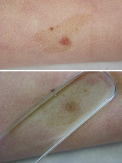
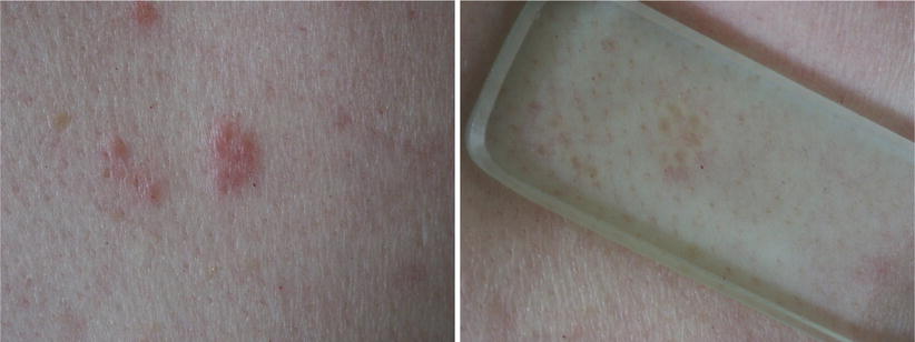
Fig. 8.1
Diascopy. Spitz Nevus. There is a red papule on a café au lait patch. The brown color of the papule appears only on diascopy. Multiple eruptive Spitz nevi can sometimes be found on café au lait patches
Fig. 8.2
Diascopy. Lupoid. Sarcoidosis. This erythematous papule turns out to be lupoid on diascopy, due to the fact that yellow (apple-jelly) grains present in the dermis become apparent when blood is expelled by the slide. This sign reflects a granulomatous disorder or a lymphoma
8.2 Wood’s Light Examination
This examination is done in the dark, under ultraviolet light (λ = 400 nm). Epidermal skin pigmentation is exacerbated, thus increasing the contrast between normal skin and depigmented areas (e.g., vitiligo, chemical leukoderma, piebaldism). This technique can also be used for the rapid detection of urine porphyrins which take on a pink color under Wood’s light, following acidification. Finally, several infectious dermatoses are revealed by their characteristic fluorescence under Wood’s light: coral red fluorescence in erythrasma, green fluorescence in dermatophytoses caused by Microsporum as well as in favus, and green-yellow fluorescence in Pseudomonas infections. When suspecting scabies, furrows can be researched under Wood’s light, after application of tetracycline or fluorescein.
8.3 Applying Certain Substances on the Skin
Useful information can be obtained:
Application of a drop of oil eliminates air in between scales and modifies the refractive index of keratin. This is especially useful in identifying Wickham’s striae of lichen.
Application of Indian ink, followed by rinsing, allows to identify scabies furrows without using a Wood’s lamp; a felt-tip pen can also be used as the coloring agent which has penetrated the furrow persists after rinsing (cf. Fig. 12.35).
8.4 Linear Stimulation
Firm stimulation using a blunt tip enables to search for dermographism (cf. Fig. 12.20). Rubbing of certain lesions elicits an urticarial response, referred to as Darier’s sign, which is characteristic of mastocytosis (cf. Figs. 15.69 and 15.70).
Related posts:


Stay updated, free articles. Join our Telegram channel

Full access? Get Clinical Tree


