8
Orbit and Zygoma Fractures
 The Orbit
Anatomy
The orbit is composed of seven bones:
• Zygoma
• Greater and lesser wing of the sphenoid
• Ethmoid
• Frontal
• Palatine
• Maxilla
• Lacrimal
These seven bones create a bony pyramid with the optic canal at the apex. The orbit is comprised of the following structures
 Floor
Floor
 Roof of the maxillary sinus
 Medial wall
 Lamina papyracea of the ethmoid bone
 Lacrimal bone
 Lateral wall
 Zygoma and greater wing of the sphenoid bone
 Roof
Roof
 Frontal bone – floor of the frontal sinus
The medial wall is the weakest structure, followed by the floor. The roof and the lateral wall are generally the strongest. The optic nerve exits the optic canal situated superomedially and ~40 to 45 mm from the inferior orbital rim. The superior orbital fissure separates the greater and lesser wings of the sphenoid. From the superior orbital fissure traverses the
• Oculomotor nerve (CN III)
• Trochlear nerve (CN IV)
• Abducens nerve (CN VI)
• Ophthalmic division of the trigeminal nerve (CN V1)
The inferior orbital fissure provides passage of the
• Maxillary division of trigeminal (CN V2)
• Branches of sphenopalatine ganglion
• Branches of the inferior ophthalmic vein
Physical Examination
Orbital fractures are usually associated with blunt trauma. Nearly 30% of orbital fractures will have injuries to the globe. It is important to perform a detailed ophthalmic exam that includes visual acuity, pupillary reaction, retinal exam, and red color saturation, as described in Chapter 7. Any deviation from normal warrants an emergent ophthalmic consultation.
Pathologic physical findings include
• Orbital ecchymosis
• Periorbital edema
• Subconjunctival hemorrhage
• Epistaxis
• Orbital rim/zygoma bony step-offs
• Diplopia
• Extraocular muscle entrapment
 Examine the active range of motion of the extraocular muscles to rule out mechanical entrapment.
 In unconscious patients, perform the forced duction test: using Adson forceps grasp the inferior capsulopalpebral fascia of the inferior rectus muscle and gently rotate the globe, while feeling for any restrictions.
• Intraorbital edema
• Optic nerve neuropraxia
• Pupillary shape – oblong pupil is suggestive of ocular perforation
• Pupillary response – afferent pupillary defect (see Chapter 7)
• Supraorbital, infraorbital, alveolar nerve paresthesias
• Crepitus/subcutaneous emphysema – disruption of maxillary or ethmoid sinus mucosa
• Enophthalmos – noticeably with >2 mm shift; however, rarely evident immediately postinjury because of edema
• Proptosis/exophthalmos
• Hyphema – fluid in the anterior chamber of the eye
• Superior orbital fissure (SOF) syndrome – fractures of the SOF result in
 Fixed dilated pupil (CN III)
 Upper lid ptosis (CN III)
 Loss of corneal reflex (CN V1)
 Ophthalmoplegia (CN IV, CN VI)
• Orbital apex syndrome – SOF syndrome plus impairment of optic nerve as it exists in the optic canal
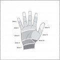
• Nausea, vomiting, bradycardia – oculocardiac response to extraocular muscle entrapment (Fig. 8–1

Stay updated, free articles. Join our Telegram channel

Full access? Get Clinical Tree


