Orbicularis Oris Muscle Flaps
C. L. PUCKETT
K. L. PICKRELL
J. F. REINISCH
Mobilization of the orbicularis oris muscle flap has been useful in reconstruction for paralysis or discontinuity of the orbicularis muscle.
INDICATIONS
The two clinical circumstances in which we have used this technique most successfully have been (a) dense paralysis of
the lip associated with injury to the marginal mandibular branch of the facial nerve (1), and (b) sphincteric discontinuity of the orbicularis oris muscle in the bilateral cleft lip patient (2).
the lip associated with injury to the marginal mandibular branch of the facial nerve (1), and (b) sphincteric discontinuity of the orbicularis oris muscle in the bilateral cleft lip patient (2).
ANATOMY
The orbicularis oris muscle is a sphincter that has no clearly defined origin and insertion; however, its lateral attachments at the modiolus can be considered the origin, with the insertion being into its contralateral half in the center of the upper and lower lips. The muscle is flattened, somewhat ribbon-like, with a thickness of approximately 0.5 cm, and it varies from 1 to 2 cm in width. It is innervated in quadrants (although there is significant overlap) by the zygomatic branches of the facial nerve for the upper half and the marginal mandibular branch of the facial nerve for the lower half (3). There is variable input by the buccal branch. Three or more filaments of these nerves may enter the muscle quadrant. Blood supply is from the facial artery by means of the labial branches.
FLAP DESIGN AND DIMENSIONS
Because of a rich blood supply, the muscle can be used as a flap based either medially or laterally. When based laterally, flap innervation can be maintained, but if based medially, the flap is effectively denervated.
Unilateral Lower Lip Paralysis
Following marginal mandibular branch injury in which nerve repair is not feasible, a medially based flap of the atrophic orbicularis oris muscle can be combined with a wedge resection of the lip to improve the appearance of the defect. This is particularly helpful when the paralysis is dense and associated with atrophy of the orbicularis oris muscle and droop of the lateral lower lip. A V-shaped wedge of lower lip is marked, with the width of the V matching the amount of orbicularis atrophy (Fig. 172.1A). The lateral limb of the V is 1 or 2 mm medial to the commissure of the lip.
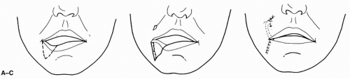 FIGURE 172.1 A: The wedge resection outlined. The lateral limb of the V is a few millimeters medial to the commissure. The approximate width of the atrophic segment indicates the width of the V. B: The wedge has been resected through and through, preserving only the atrophic strand of orbicularis oris. Note the incision in the nasolabial fold above the commissure. C: The wedge has been approximated, and the orbicularis oris muscle flap has been brought through the subcutaneous tunnel and sutured to the region of the insertion of the zygomaticus major. (From Puckett et al., ref. 1, with permission.)
Related posts:Stay updated, free articles. Join our Telegram channel
Full access? Get Clinical Tree
 Get Clinical Tree app for offline access

|


