Fig. 2.1
Surface anatomy demonstrating the relations between the nipple and areola complex, the sternum, and the inframammary fold
2.4 Surgical Anatomy of the Breast
The breasts are located over the pectoralis major muscle. Considering their structure, of cutaneous origin, a layer of adipose tissue and a layer of mammary tissue form them. The plane that separates the mammary tissue from the adipose tissue has great surgical value, as it can be identified in the flap during the surgical procedure and can be utilized in mastectomies and even in conservative surgical procedures.
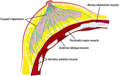
In relation to the covering of the mammary tissue by fascia, it is important to consider the superficial fascia, which is found over the deep fascia of the skin (2–3 mm below), except at the areola and the nipple. This layer can be identified during the surgical dissection, allowing the separation of the mammary tissue from the skin where there is no bleeding, in an avascular plane. This fascia is connected with the deep fascia of the breast through fibrous fascia called Cooper’s ligaments, which support the breast. On the posterior face of the gland there is a layer of thin adipose tissue that connects with the superficial fascia. This is separated from the pectoralis major muscle fascia by a layer of dense connective tissue called the posterior suspensory ligament of the breast. At the lateral and the inferior borders the breast lies on the fascia of the anterior serratus muscle and the anterior rectus abdominis muscle, respectively [6, 9].
Fifteen to twenty independent lobes that branch out in lobules and alveoli form the mammary gland. A lobe is made up of various lobules that branch out in ten to 12 alveoli. These form the functional units of the breast, and are responsible for the production and drainage of milk. They are enveloped by fatty fibrous tissue. The drainage system is made up of collecting ducts that drain to the lactiferous ducts situated at a retroareolar position where milk is stored and ejected then by the apex of the nipple [6] (Fig. 2.2).


Fig. 2.2
Relationship between breast lobes, Cooper’s ligaments, fat tissue, and thoracic muscles
The subclavian artery, the axillary artery, and intercostal arteries and their branches form the arterial vascularization of the breast. The knowledge of their relationships is important, as in reduction mammoplasties. It is essential that at least one of these arterial pathways be preserved so the areola and the nipple can remain viable. Therefore, there are techniques for the preservation of the upper pedicle, the lateral pedicle, or the inferior pedicle blood supplies.
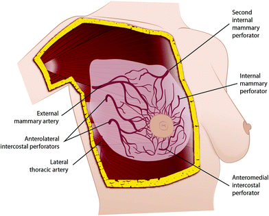
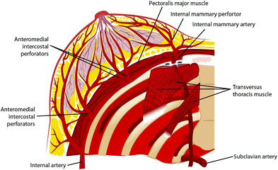
The breast is supplied with blood coming from three main sources. The first one and also the most important one (representing over 60 % of the supply) is the internal mammary artery, which may originate from the second, third, or fourth intercostal space. It perforates these spaces and enters the breast in its superomedial portion, taking a superficial track from where it sends arterioles to the skin and to the mammary tissue [5, 6, 9]. It is important to identify and preserve the integrity of this system while making the skin flaps during SSM.
The second system originates in the axillary artery. The pectoral branch of the thoracoacromial artery, the highest thoracic artery, subscapular artery, and mainly the lateral mammary branches of the lateral thoracic artery are responsible for about 30 % of the blood supply of the breast. One of the lateral mammary branches is more developed and mainly supplies the upper outer quadrant of the breast [5, 6, 9]. The surgical relevance of this vessel is because it is used for mammary reconstruction procedures that demand microsurgical anastomoses.
The third source of breast blood supply comes from lateral cutaneous branches of the intercostal arteries, which are less important [5, 6, 9] (Figs. 2.3, 2.4).


Fig. 2.3
Oblique view of the arterial vascularization of the breast

Fig. 2.4
Coronal view of the arterial vascularization of the breast
The venous drainage is formed by a deep system and a superficial system. Low-caliber vessels that drain just below the superficial fascia form an interconnected traverse longitudinal network, like a knit cloth, which drains to the internal mammary vein and the anterior superficial jugular vein, the superficial system. In the deep system, the afferent branches discharge into three main pathways: tributaries of the internal mammary vein, tributaries of the intercostal veins, and the vertebral system. There is special interest in the mammary venous drainage owing to the potential use of certain branches in breast reconstructive surgical procedures. The drainage follows the course of the arteries, with a large number of anastomoses between the superficial and the deep system, and has the axillary vein (originating from the cephalic vein and the humeral vein) as its main system. Around the areola, the veins form a venous circle which together with the drainage of the mammary tissue follows a peripheral course up to the internal thoracic, axillary, and intercostal veins [6, 9]. Metastases can pass through any of these routes, following their way to the heart and then to the lung capillaries. Owing to a system of avalvular venous drainage that connects Batson’s venous vertebral plexus to thoracic, abdominal, and pelvic organs, one can explain the route of metastases to the vertebra, ribs, and central nervous system from the breast, mainly through intercostal posterior veins.
Interest in studying lymphatic drainage has increased because of SN studies. In most cases, the SN position is at level I, close to the thoracodorsal artery and vein. The breast lymph vessels have their drainage established by two plexuses: superficial or Sappey’s subareolar, and deep or aponeurotic. The former is made up by collecting trunks, which gather skin drainage, superficial breast planes, the nipple and areola, and the upper limb, supraumbilical region, and dorsum. The latter follows through the pectoralis muscles up to Rotter’s lymph nodes (situated between the pectoralis major and the pectoralis minor muscles) and then toward the subclavian lymph nodes. Although the lymphatic flow is unidirectional, there is a great interrelation between the superficial system and the deep system as to breast drainage, which explains the broad variation of lymph drainage found in breast cancer [12]. Approximately 3 % of the breast lymph flows to the lymph nodes in the internal mammary chain and 97 % flows to the axillary lymph nodes. Any quadrant of the breast is able to drain to the internal mammary chain. Axillary nodes range in number from 20 to 30, and lymph node groups of axillary drainage can be divide into [5]:
The axillary vein group or lateral group, consisting of four to six lymph nodes located medial or posterior to the axillary vein, holding most of the drainage from the superior portion of the breast.
The external mammary group, also called the pectoral group, situated at the inferior border of the pectoralis minor muscle in association with the lateral thoracic vessels. It consists of four or five lymph nodes and holds most of the lymphatic drainage from the breast.
The subscapular lymph node group or posterior lymph node group, consisting of six or seven lymph nodes situated along the posterior wall of the axilla up to the lateral border of the scapula and are associated with subscapular vessels. They also contain drainage from the cervical posterior region and the shoulder.
The central group, consisting of three or four lymph nodes and situated posterior to the pectoralis minor muscle, interwoven with the adipose tissue of the axilla. They hold drainage from the three groups mentioned above and they can also contain drainage directly from the breast. A sequence of this drainage moves on to the subclavicular lymph nodes or to apical lymph nodes. Clinically, this is the most palpable group, which is of extreme relevance to the clinical evaluation of axillary metastases.
The subclavicular or apical group, consisting of six to 12 lymph nodes, situated posterior and superior to the border of the pectoralis minor muscle. It obtains drainage directly or indirectly from all the other groups. The lymphatic efferents of these ducts form the subclavian trunk, which pours into the right lymphatic duct and to the left side into the thoracic duct. Through this route there is also the possibility of drainage for lymph nodes from the deep cervical area.
Rotter’s group or the interpectoral group, consisting of one to four small lymph nodes situated between the pectoralis major and the pectoralis minor muscles associated with branches of thoracoacromial vessels.
Stay updated, free articles. Join our Telegram channel

Full access? Get Clinical Tree


