22.1 Non-vascularized Nerve Graft (Level of Difficulty: 3)
Information
There are various reports on locations where nerves can be harvested, however the following three types are practical with small loss from the donor site. Indication varies according to the thickness of the reconstructed nerve
- 1.
Cutaneous nerve of the forearm
- 2.
Posterior interosseous nerve
- 3.
Sural nerve
For fingers, either the cutaneous nerve of the forearm or the posterior interosseous nerve is suitable; for the palm area, the sural nerve is more practical. In order to reconstruct a thick nerve, several thin nerves are bundled together and used as a cable graft

22.1.1 Operation Procedures
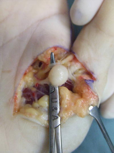
Case 1Fig. 22.1
Case 1: Procedure 1: An amputation neuroma is present on the radial digital nerve of the thumb
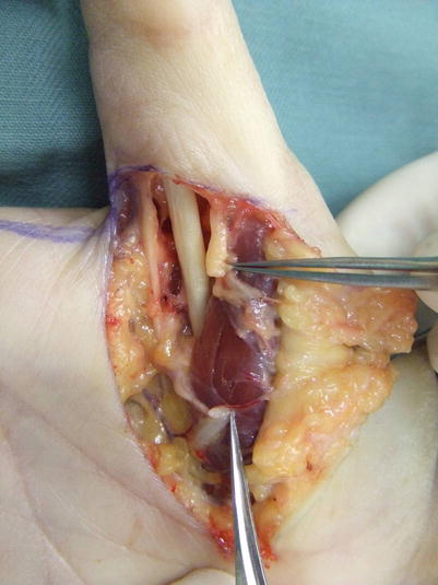
Fig. 22.2
Procedure 2: Due to resection of the neuroma, a loss of around 20 mm of nerve occurred

Fig. 22.3
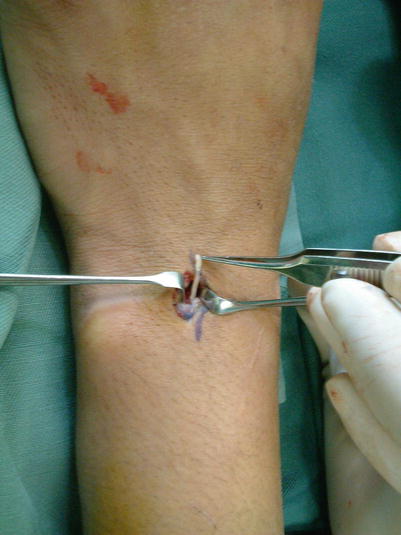
Procedure 3: A small incision is made in the middle of the medial forearm, and after detaching the antebrachial fascia, it becomes possible to confirm the thin cutaneous nerve of the forearm. The required length of this nerve is severed and harvested
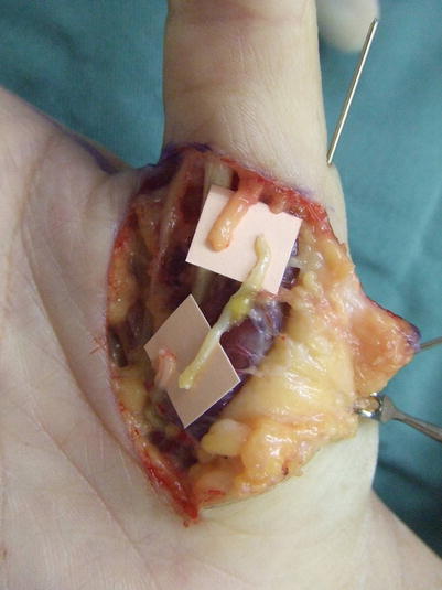
Fig. 22.4
Procedure 4: The proximal and distal ends of the harvested cutaneous nerve of the forearm are reversed and transferred to the thumb
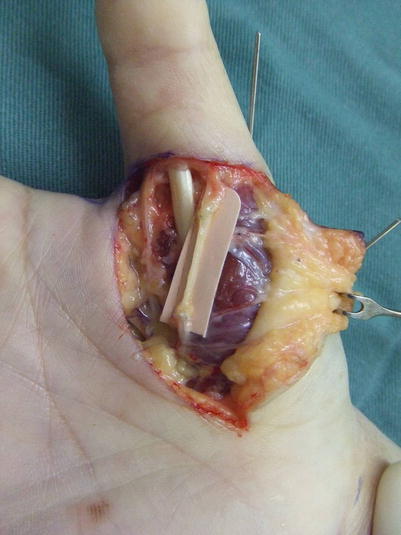
Fig. 22.5
Procedure 5: It is sutured under an optical microscope using a 9-0, 10-0 nylon suture
Case 2Fig. 22.6
Case 2: Procedure 1: Harvesting of posterior interosseous nerve: A small incision is made beneath the 4th compartment of the dorsal wrist joint, enabling confirmation of the posterior interosseous nerve rising up between the radius and the ulna. Around 20 mm of this 1 mm diameter nerve can be harvested
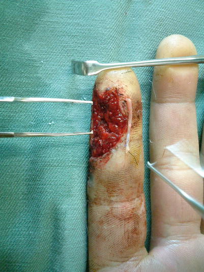
Fig. 22.7
Procedure 2: This can be used for the reconstruction of the finger nerves of the distal fingertips

Case 3Fig. 22.8
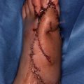
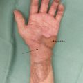
Case 3: Procedure 1: A 2 cm nerve defect was present following resection of an amputation neuroma on the median nerve on the left hand

Fig. 22.9
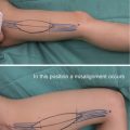
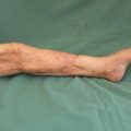
Procedure 2: An incision is made to the left lateral Achilles tendon above the posterosuperior lateral malleolus, enabling confirmation of the sural nerve running beneath the subcutaneous fat. The required amount is harvested
Note
One method of harvesting this involves making multiple small horizontal incisions every few centimeters, but because it is easily damaged, the author chooses to harvest using a longitudinal incision.
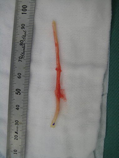
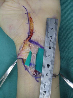
Fig. 22.10
Procedure 3: Because the sural nerve is thin, when transferring to the median nerve, several lengths are bundled together for use as a cable graft. Therefore it is necessary to harvest the necessary length of sural nerve, with care taken that the distal end is marked and sutured to the proximal end of the median nerve
Fig. 22.11


Procedure 4: Three sural nerves are bundled together and a cable graft is conducted
Related posts:
Stay updated, free articles. Join our Telegram channel
Full access? Get Clinical Tree
