Nasolabial Dermal Flaps for Suspension in Lower Facial Palsy
M. R. WEXLER
I. J. PELED
EDITORIAL COMMENT
This chapter presents a helpful adjunct in restoration of facial symmetry. The procedure can be significantly augmented by the addition of fascia lata slings connected to the temporalis muscle.
INDICATIONS
Patients are often interested in a simpler procedure and reject the idea of an operation on the normal side of the face or elsewhere on the body. We offer our patients a variety of procedures, and our experience is based on those who objected to more sophisticated techniques.
For several years, we have used dermal nasolabial flaps for suspension to improve lower facial palsy (4, 5). The advantages of this method are its simplicity, the use of tissue otherwise discarded, and the creation of a fibrotic cheek that improves speech. The nasolabial fold is simulated by the new scar. Correction of lower eyelid ectropion can be achieved at the same time, although the results are only average. Elevation of redundant ala nasi can be achieved as well. If the results are not satisfactory, further procedures can be performed.
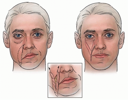
 FIGURE 152.1 Schematic drawing of the procedure. Note that the lateral flap is elongated in the lower part. (From Wexler et al., ref. 5, with permission.) |
FLAP DESIGN AND DIMENSIONS
A fusiform area is marked along the nasolabial fold of the affected side (Figs. 152.1 and 152.2B) from the upper pole of the ala nasi to the border of the mandible. The average size of the flap is 9 cm long and 3 cm wide. The rich blood supply
of the nasolabial area allows the raising of quite long flaps that are narrowly based at the corner of the mouth.
of the nasolabial area allows the raising of quite long flaps that are narrowly based at the corner of the mouth.
Stay updated, free articles. Join our Telegram channel

Full access? Get Clinical Tree


