Nail procedures require an effective and reliable approach to anesthesia of the distal digit. Several techniques have been described in the literature. Herein, the relevant anatomy of the nail unit, pain pathways, anesthetic options, and several injection approaches to achieve complete anesthesia are reviewed. Also considered are the potential pitfalls and complications and their management. Ultimately, the physician’s approach must be individualized to the patient, procedure, and setting.
Key points
- •
Anesthesia of the nail unit requires a complete understanding of the anatomic and physiologic pathways of pain and the different anesthetic choices.
- •
Buffering and warming the local anesthetic coupled with a slow rate of injection and small needle size, all drastically reduce pain of injection.
- •
Local infiltrative anesthesia, termed a wing block, is an efficient and well-tolerated form of anesthesia; however, proper performance uses distracting anesthesia and slow rate of injection.
- •
Traditional digital block involves injections at the base/sides of the digit and allowance of time for anesthesia to take effect.
- •
Single-digit injection techniques (transthecal) are effective on the second to fourth fingers and provide complete anesthesia; however, postoperative pain may be more than with other techniques.
Introduction, treatment goals, and planned outcomes
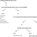
Successful nail surgery requires complete anesthesia. Sometimes it is this initial step in nail surgery that most intimidates the patient and, quite often, the physician. As such, mastery of digital anesthesia is a prerequisite to performing competent surgery on the nail apparatus. Administering digital anesthesia requires a multifaceted understanding of the anatomy of the digit, pathophysiology of pain, mechanisms of local anesthetics, and nuances in both technique and preparation that maximize effectiveness of the procedure.
Digital nerves run along each digit as paired parallel volar and dorsal nerves, terminating just beyond the distal interphalangeal joint (DIPJ), where they divide into 3 branches, supplying the nail bed, the digital tip, and pulp. There is no clear consensus on which specific branches innervate the tips for each digit. Generally accepted dogma is that the second to fourth fingertips are innervated by the volar branches, whereas the thumb and fifth fingertips are innervated primarily by the dorsal branches. These sensory nerves carry the impulses from the many smaller nociceptors located in the nail unit and surrounding tissue to the brain.
Cutaneous nociceptors provide an innate protective warning system for injury and consist of Pacinian corpuscles (movement-sensitive hair follicle receptors), Ruffini corpuscles (pressure sensitive mechanoreceptors), and free-ended nociceptors located at the dermoepidermal junction. There are 2 main classes of nerve fibers—fast/myelinated (A-δ, carrying sharp pain) and slow/unmyelinated (C, carrying dull pain), which are activated by these receptors and transfer impulses of pain. Local anesthetics work by blocking the free nerve endings’ voltage-gated sodium channels and nerve depolarization, thus impeding transmission of pain. However, local anesthetics must first diffuse into the nerve cells through hydrophobic cell membranes.
During anesthesia, patients experience pain from 2 distinct and unrelated procedures—the needle insertion and fluid infiltration. The former activates Pacinian corpuscles and mechanoreceptors, which transmit via A-δ fibers to evoke the pinprick sensation, whereas the latter (through chemical irritation and rapid distention of tissue) activates mostly free-ending nociceptors and produces a more intense and continuous pain. Infiltrative anesthesia results from anesthesia of the smallest nerve fibers and blocking initial transmission of nociception, whereas nerve blocks affect larger, usually more proximal nerve fibers and require longer time of onset to diffuse into the nerves and block depolarization.
Introduction, treatment goals, and planned outcomes
Successful nail surgery requires complete anesthesia. Sometimes it is this initial step in nail surgery that most intimidates the patient and, quite often, the physician. As such, mastery of digital anesthesia is a prerequisite to performing competent surgery on the nail apparatus. Administering digital anesthesia requires a multifaceted understanding of the anatomy of the digit, pathophysiology of pain, mechanisms of local anesthetics, and nuances in both technique and preparation that maximize effectiveness of the procedure.
Digital nerves run along each digit as paired parallel volar and dorsal nerves, terminating just beyond the distal interphalangeal joint (DIPJ), where they divide into 3 branches, supplying the nail bed, the digital tip, and pulp. There is no clear consensus on which specific branches innervate the tips for each digit. Generally accepted dogma is that the second to fourth fingertips are innervated by the volar branches, whereas the thumb and fifth fingertips are innervated primarily by the dorsal branches. These sensory nerves carry the impulses from the many smaller nociceptors located in the nail unit and surrounding tissue to the brain.
Cutaneous nociceptors provide an innate protective warning system for injury and consist of Pacinian corpuscles (movement-sensitive hair follicle receptors), Ruffini corpuscles (pressure sensitive mechanoreceptors), and free-ended nociceptors located at the dermoepidermal junction. There are 2 main classes of nerve fibers—fast/myelinated (A-δ, carrying sharp pain) and slow/unmyelinated (C, carrying dull pain), which are activated by these receptors and transfer impulses of pain. Local anesthetics work by blocking the free nerve endings’ voltage-gated sodium channels and nerve depolarization, thus impeding transmission of pain. However, local anesthetics must first diffuse into the nerve cells through hydrophobic cell membranes.
During anesthesia, patients experience pain from 2 distinct and unrelated procedures—the needle insertion and fluid infiltration. The former activates Pacinian corpuscles and mechanoreceptors, which transmit via A-δ fibers to evoke the pinprick sensation, whereas the latter (through chemical irritation and rapid distention of tissue) activates mostly free-ending nociceptors and produces a more intense and continuous pain. Infiltrative anesthesia results from anesthesia of the smallest nerve fibers and blocking initial transmission of nociception, whereas nerve blocks affect larger, usually more proximal nerve fibers and require longer time of onset to diffuse into the nerves and block depolarization.
Preoperative planning and preparation, patient positioning, “best way to perform”
Several factors may impact the patient’s degree of pain from anesthesia and their postoperative discomfort. These factors include both specific characteristics of the anesthetic (molecular composition, pH, temperature, addition of epinephrine) and choice of syringe and needle size, distracting stimuli, and technique of injection. Each of these considerations is highlighted individually below.
Three main anesthetics are used in digital anesthesia—lidocaine, bupivacaine, and ropivacaine ( Tables 1 and 2 ). Lidocaine is still the most widely used and has an unparalleled safety history. It is estimated that more than 300 million doses of lidocaine with epinephrine are injected in dental offices alone each year in the United States. Lidocaine is characterized by quick absorption and near instantaneous anesthesia of the minute nociceptors in the skin. The onset is faster (<1–3 min) than that of either ropivacaine (4.5 min) or bupivacaine (4+ min), with a significantly shorter duration of action (60–120 min). Bupivacaine has a longer onset of action and a longer duration of action (480 min). Ironically, a combination of lidocaine and bupivacaine has not reliably shown superiority over each agent alone. Ropivacaine represents perhaps the current ideal in terms of anesthetic, with a short onset of action, prolonged duration of action (up to 20 hours) and less cardiotoxicity than bupivacaine. In addition, ropivacaine has demonstrated some degree of inherent vasoconstriction (as opposed to the vasodilating effect of lidocaine), with potential benefits of providing a bloodless field. A down side to consider with the longer acting anesthetics is the risk of masking pain associated with postoperative complications, including compartment syndrome, infection, and ischemia.
| Anesthetic | Onset (min) | pKa | Duration Without Epinephrine | Duration with Epinephrine | Benefits |
|---|---|---|---|---|---|
| Lidocaine | <1 | 7.7 | 30–120 | 60–400 | Near instantaneous onset |
| Bupivacaine | 2–5 | 8.1 | 120–240 | 240–480 | Longer duration |
| Ropivacaine | 1–15 | 8.2 | 120–360 | Not defined | Longer duration, Potential vasoconstrictive effects |
| Technique | Considerations | Pros | Cons | |
|---|---|---|---|---|
| Infiltrative | ||||
| Wing Block | Inject wheal (0.1–0.2 ml) in proximal nail fold and then inject slowly toward each lateral nail fold to digital tip. | Avoid rapid tissue distention. Most anesthesia should be on dorsal aspect of digit. | Near instantaneous anesthesia. Hemostasis from tissue tamponade. | Discomfort without distracting stimuli or with overzealous injection. Potential for tissue tourniquet if too much fluid injected into pulp. |
| Nerve Blocks | ||||
| TDB | Insert needle into the web space at the level of MCP/MTP joint and inject into the subcutaneous tissue. | During delay of anesthesia, consider soaking digit in chlorhexidine and water. | More complete anesthesia of entire digit. More prolonged anesthesia. | Not instantaneous. Requires (at least) 2 needle punctures. May still require infiltrative anesthesia. |
| TTB | Insert needle in to the palmar digital creased to bone and inject on pullback, as needle tip exits tendon (within tendon sheath). | Requires 3 ml of anesthesia. Can also inject in same location subcutaneously followed by massage (modified subcutaneous technique). | Single injection | Potentially more postoperative pain at injection site. |
Related posts:


Stay updated, free articles. Join our Telegram channel

Full access? Get Clinical Tree


